 Movie
Movie Controller
 Structure viewers
 About Yorodumi Papers


+Search query
-Structure paper
| Title | Cryo-EM structures of S-OPA1 reveal its interactions with membrane and changes upon nucleotide binding. |
|---|---|
| Journal, issue, pages | Elife, Vol. 9, Year 2020 |
| Publish date | Mar 31, 2020 |
 Authors Authors | Danyang Zhang / Yan Zhang / Jun Ma / Chunmei Zhu / Tongxin Niu / Wenbo Chen / Xiaoyun Pang / Yujia Zhai / Fei Sun /  |
| PubMed Abstract | Mammalian mitochondrial inner membrane fusion is mediated by optic atrophy 1 (OPA1). Under physiological conditions, OPA1 undergoes proteolytic processing to form a membrane-anchored long isoform (L- ...Mammalian mitochondrial inner membrane fusion is mediated by optic atrophy 1 (OPA1). Under physiological conditions, OPA1 undergoes proteolytic processing to form a membrane-anchored long isoform (L-OPA1) and a soluble short isoform (S-OPA1). A combination of L-OPA1 and S-OPA1 is essential for efficient membrane fusion; however, the relevant mechanism is not well understood. In this study, we investigate the cryo-electron microscopic structures of S-OPA1-coated liposomes in nucleotide-free and GTPγS-bound states. S-OPA1 exhibits a general dynamin-like structure and can assemble onto membranes in a helical array with a dimer building block. We reveal that hydrophobic residues in its extended membrane-binding domain are critical for its tubulation activity. The binding of GTPγS triggers a conformational change and results in a rearrangement of the helical lattice and tube expansion similar to that of S-Mgm1. These observations indicate that S-OPA1 adopts a dynamin-like power stroke membrane remodeling mechanism during mitochondrial inner membrane fusion. |
 External links External links | Elife / PubMed:32228866 / PubMed Central |
| Methods | EM (subtomogram averaging) / EM (helical sym.) |
| Resolution | 15.0 - 31.7 Å |
| Structure data |  EMDB-0722:  EMDB-9901: 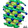 EMDB-9902:  EMDB-9903: |
| Source |
|
 Homo sapiens (human)