 ムービー
ムービー コントローラー


+ データを開く

- 基本情報
基本情報
| 登録情報 | データベース: EMDB / ID: EMD-3724 | |||||||||
|---|---|---|---|---|---|---|---|---|---|---|
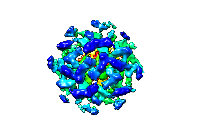
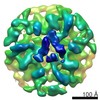
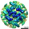
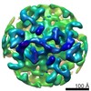
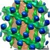
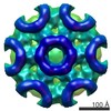
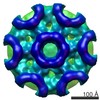
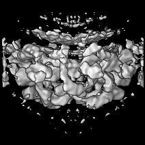
| タイトル | The structure of the COPI coat linkage IV | |||||||||
 マップデータ マップデータ | The structure of the COPI coat linkage IV | |||||||||
 試料 試料 |
| |||||||||
 キーワード キーワード | COPI / coatomer / coated vesicles / Transport protein | |||||||||
| 機能・相同性 |  機能・相同性情報 機能・相同性情報cerebellar Purkinje cell layer maturation / Synthesis of PIPs at the plasma membrane / protein localization to axon / VxPx cargo-targeting to cilium / protein localization to cell leading edge / Synthesis of PIPs at the Golgi membrane / protein localization to Golgi membrane / Golgi localization / regulation of Golgi organization / COPI-coated vesicle ...cerebellar Purkinje cell layer maturation / Synthesis of PIPs at the plasma membrane / protein localization to axon / VxPx cargo-targeting to cilium / protein localization to cell leading edge / Synthesis of PIPs at the Golgi membrane / protein localization to Golgi membrane / Golgi localization / regulation of Golgi organization / COPI-coated vesicle / pancreatic juice secretion / trans-Golgi Network Vesicle Budding / organelle membrane contact site / Intra-Golgi traffic / COPI vesicle coat / COPI-mediated anterograde transport / Golgi vesicle transport / COPI-dependent Golgi-to-ER retrograde traffic / COPI-mediated anterograde transport / positive regulation of mitochondrial fusion / COPI-dependent Golgi-to-ER retrograde traffic / organelle transport along microtubule / regulation of fatty acid metabolic process / establishment of Golgi localization / Golgi to plasma membrane transport / intra-Golgi vesicle-mediated transport / retrograde vesicle-mediated transport, Golgi to endoplasmic reticulum / positive regulation of mitochondrial fission / Golgi-associated vesicle / endoplasmic reticulum-Golgi intermediate compartment / pigmentation / protein secretion / endoplasmic reticulum to Golgi vesicle-mediated transport / vesicle-mediated transport / Neutrophil degranulation / protein kinase C binding / small monomeric GTPase / adult locomotory behavior / macroautophagy / hormone activity / intracellular protein transport / protein transport / growth cone / cell body / G protein activity / Golgi membrane / axon / mRNA binding / GTPase activity / GTP binding / structural molecule activity / Golgi apparatus / endoplasmic reticulum / : / nucleoplasm / plasma membrane / cytoplasm / cytosol 類似検索 - 分子機能 | |||||||||
| 生物種 |   | |||||||||
| 手法 | サブトモグラム平均法 / クライオ電子顕微鏡法 / 解像度: 17.3 Å | |||||||||
 データ登録者 データ登録者 | Dodonova SO / Aderhold P | |||||||||
| 資金援助 |  ドイツ, 2件 ドイツ, 2件
| |||||||||
 引用 引用 | ジャーナル: Elife / 年: 2017 タイトル: 9Å structure of the COPI coat reveals that the Arf1 GTPase occupies two contrasting molecular environments. 著者: Svetlana O Dodonova / Patrick Aderhold / Juergen Kopp / Iva Ganeva / Simone Röhling / Wim J H Hagen / Irmgard Sinning / Felix Wieland / John A G Briggs /  要旨: COPI coated vesicles mediate trafficking within the Golgi apparatus and between the Golgi and the endoplasmic reticulum. Assembly of a COPI coated vesicle is initiated by the small GTPase Arf1 that ...COPI coated vesicles mediate trafficking within the Golgi apparatus and between the Golgi and the endoplasmic reticulum. Assembly of a COPI coated vesicle is initiated by the small GTPase Arf1 that recruits the coatomer complex to the membrane, triggering polymerization and budding. The vesicle uncoats before fusion with a target membrane. Coat components are structurally conserved between COPI and clathrin/adaptor proteins. Using cryo-electron tomography and subtomogram averaging, we determined the structure of the COPI coat assembled on membranes in vitro at 9 Å resolution. We also obtained a 2.57 Å resolution crystal structure of βδ-COP. By combining these structures we built a molecular model of the coat. We additionally determined the coat structure in the presence of ArfGAP proteins that regulate coat dissociation. We found that Arf1 occupies contrasting molecular environments within the coat, leading us to hypothesize that some Arf1 molecules may regulate vesicle assembly while others regulate coat disassembly. | |||||||||
| 履歴 |
|
- 構造の表示
構造の表示
| ムービー |
ムービービューア |
|---|---|
| 構造ビューア | EMマップ: SurfViewMolmilJmol/JSmol |
| 添付画像 |
- ダウンロードとリンク
-EMDBアーカイブ
| マップデータ | emd_3724.map.gz | 33.8 MB | EMDBマップデータ形式 | |
|---|---|---|---|---|
| ヘッダ (付随情報) | emd-3724-v30.xmlemd-3724.xml | 27.9 KB 27.9 KB | 表示 表示 | EMDBヘッダ |
| FSC (解像度算出) | emd_3724_fsc.xml | 8.1 KB | 表示 | FSCデータファイル |
| 画像 |  emd_3724.png emd_3724.png | 127.2 KB | ||
| Filedesc metadata | emd-3724.cif.gz | 10.2 KB | ||
| アーカイブディレクトリ |  http://ftp.pdbj.org/pub/emdb/structures/EMD-3724ftp://ftp.pdbj.org/pub/emdb/structures/EMD-3724 http://ftp.pdbj.org/pub/emdb/structures/EMD-3724ftp://ftp.pdbj.org/pub/emdb/structures/EMD-3724 | HTTPS FTP |
-関連構造データ
| 関連構造データ | 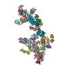 5nzvMC 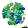 3720C 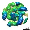 3721C 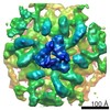 3722C 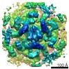 3723C 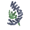 5mu7C 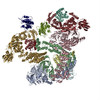 5nzrC 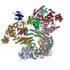 5nzsC 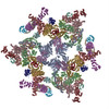 5nztC 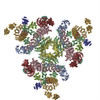 5nzuC M: このマップから作成された原子モデル C: 同じ文献を引用 ( |
|---|---|
| 類似構造データ |
-リンク
| EMDBのページ | EMDB (EBI/PDBe) / EMDataResource |
|---|---|
| 「今月の分子」の関連する項目 |


-マップ
| ファイル | ダウンロード / ファイル: emd_3724.map.gz / 形式: CCP4 / 大きさ: 36.3 MB / タイプ: IMAGE STORED AS FLOATING POINT NUMBER (4 BYTES) | ||||||||||||||||||||||||||||||||||||||||||||||||||||||||||||
|---|---|---|---|---|---|---|---|---|---|---|---|---|---|---|---|---|---|---|---|---|---|---|---|---|---|---|---|---|---|---|---|---|---|---|---|---|---|---|---|---|---|---|---|---|---|---|---|---|---|---|---|---|---|---|---|---|---|---|---|---|---|
| 注釈 | The structure of the COPI coat linkage IV | ||||||||||||||||||||||||||||||||||||||||||||||||||||||||||||
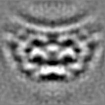
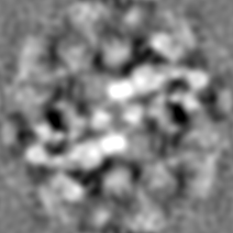
| 投影像・断面図 | 画像のコントロール
画像は Spider により作成 | ||||||||||||||||||||||||||||||||||||||||||||||||||||||||||||
| ボクセルのサイズ | X=Y=Z: 1.78 Å | ||||||||||||||||||||||||||||||||||||||||||||||||||||||||||||
| 密度 |
| ||||||||||||||||||||||||||||||||||||||||||||||||||||||||||||
| 対称性 | 空間群: 1 | ||||||||||||||||||||||||||||||||||||||||||||||||||||||||||||
| 詳細 | EMDB XML:
CCP4マップ ヘッダ情報:
| ||||||||||||||||||||||||||||||||||||||||||||||||||||||||||||
 Z (Sec.)
 Y (Row.)
 X (Col.)


-添付データ
- 試料の構成要素
+全体 : The structure of the COPI coat leaf
+超分子 #1: The structure of the COPI coat leaf
+超分子 #2: COPI coat complex
+超分子 #3: ADP-ribosylation factor 1
+分子 #1: Coatomer subunit alpha

 Spodoptera frugiperda (ツマジロクサヨトウ)
+分子 #2: Coatomer subunit epsilon
+分子 #3: Coatomer subunit beta
+分子 #4: Coatomer subunit beta'
+分子 #5: Coatomer subunit delta
+分子 #6: ADP-ribosylation factor 1

+分子 #7: Coatomer subunit gamma-1
+分子 #8: Coatomer subunit zeta-1
-実験情報
-構造解析
| 手法 | クライオ電子顕微鏡法 |
|---|---|
 解析 解析 | サブトモグラム平均法 |
| 試料の集合状態 | 3D array |
-試料調製
| 緩衝液 | pH: 7.4 構成要素:
詳細: Protein-A conjugated 10 nm gold was added to the reaction mix in 1:6 volume ratio before plunge-freezing | ||||||||||||
|---|---|---|---|---|---|---|---|---|---|---|---|---|---|
| グリッド | モデル: C-flat / 材質: COPPER / メッシュ: 200 / 支持フィルム - 材質: CARBON / 支持フィルム - トポロジー: HOLEY / 前処理 - タイプ: GLOW DISCHARGE / 前処理 - 時間: 30 sec. / 前処理 - 雰囲気: AIR / 詳細: Protochips C-flat MultiHole 20 mA | ||||||||||||
| 凍結 | 凍結剤: ETHANE / チャンバー内湿度: 85 % / チャンバー内温度: 296 K / 装置: HOMEMADE PLUNGER 詳細: The sample was applied onto glow-discharged (30 sec, 20 mA) C-flat (Protochips Inc.) multihole grids. The grids were blotted from the back side for 11 seconds at room temperature in a chamber ...詳細: The sample was applied onto glow-discharged (30 sec, 20 mA) C-flat (Protochips Inc.) multihole grids. The grids were blotted from the back side for 11 seconds at room temperature in a chamber at 85% humidity and plunge-frozen into liquid ethane using a manual plunger.. | ||||||||||||
| 詳細 | COPI-coated vesicles were produced in vitro by incubating coatomer, Arf1, GTPgS, ARNO and GUVs in a total volume of 40 ul for 30 minutes at 37C |
- 電子顕微鏡法
| 顕微鏡 | FEI TITAN KRIOS |
|---|---|
| 特殊光学系 | エネルギーフィルター - 名称: GIF Quantum LS エネルギーフィルター - エネルギー下限: 0 eV エネルギーフィルター - エネルギー上限: 20 eV |
| 詳細 | Tomographic tilt series were acquired with the dose-symmetric tilt-scheme (Hagen et al., J Struct Biol. 2017) |
| 撮影 | フィルム・検出器のモデル: GATAN K2 QUANTUM (4k x 4k) 検出モード: SUPER-RESOLUTION / デジタル化 - 画像ごとのフレーム数: 1-5 / 撮影したグリッド数: 1 / 平均電子線量: 2.0 e/Å2 詳細: Each of the images in the tilt series was low-pass filtered according to the electron-dose acquired by the sample (Grant and Grigorieff, 2015). |
| 電子線 | 加速電圧: 300 kV / 電子線源:  FIELD EMISSION GUN FIELD EMISSION GUN |
| 電子光学系 | 照射モード: FLOOD BEAM / 撮影モード: BRIGHT FIELD / Cs: 2.7 mm / 最大 デフォーカス(公称値): 5.0 µm / 最小 デフォーカス(公称値): 2.5 µm |
| 試料ステージ | 試料ホルダーモデル: FEI TITAN KRIOS AUTOGRID HOLDER ホルダー冷却材: NITROGEN |
| 実験機器 |  モデル: Titan Krios / 画像提供: FEI Company |
-画像解析
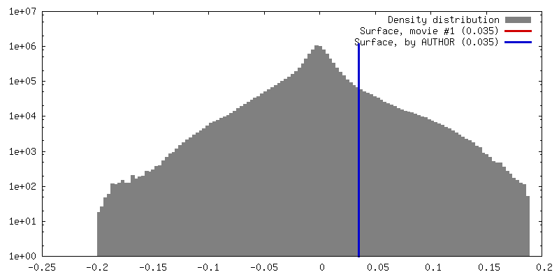
| 最終 再構成 | 想定した対称性 - 点群: C2 (2回回転対称) / 解像度のタイプ: BY AUTHOR / 解像度: 17.3 Å / 解像度の算出法: FSC 0.143 CUT-OFF / ソフトウェア: (名称: TOM Toolbox, AV3) / 使用したサブトモグラム数: 1640 |
|---|---|
| 抽出 | トモグラム数: 54 / 使用した粒子像数: 1640 / ソフトウェア: (名称: Amira, TOM Toolbox) 詳細: 1733 vesicles and near-complete buds were picked from 61 tomograms. Subtomograms were extracted from the surface of the vesicles. |
| 最終 角度割当 | タイプ: NOT APPLICABLE |
| FSC曲線 (解像度の算出) | 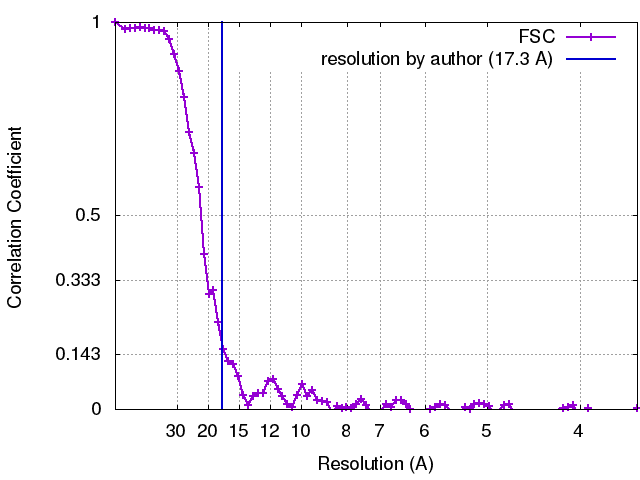 |
-原子モデル構築 1
| 精密化 | 空間: REAL / プロトコル: RIGID BODY FIT 当てはまり具合の基準: Cross-correlation coefficient |
|---|---|
| 得られたモデル | PDB-5nzv: |
