National Institutes of Health/National Institute of General Medical Sciences (NIH/NIGMS)
R01GM08139
United States
National Institute of Health Midwest Consortium for High Resolution Cryo-electron Microscopy
U24 GM116789-01A1
United States
National Science Foundation (NSF, United States)
Eager 1558128
United States
Citation
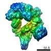
Journal: Proc Natl Acad Sci U S A / Year: 2019 Title: Structural and functional analyses of photosystem II in the marine diatom . Authors: Orly Levitan / Muyuan Chen / Xuyuan Kuang / Kuan Yu Cheong / Jennifer Jiang / Melissa Banal / Nikhita Nambiar / Maxim Y Gorbunov / Steven J Ludtke / Paul G Falkowski / Wei Dai / Abstract: A descendant of the red algal lineage, diatoms are unicellular eukaryotic algae characterized by thylakoid membranes that lack the spatial differentiation of stroma and grana stacks found in green ...A descendant of the red algal lineage, diatoms are unicellular eukaryotic algae characterized by thylakoid membranes that lack the spatial differentiation of stroma and grana stacks found in green algae and higher plants. While the photophysiology of diatoms has been studied extensively, very little is known about the spatial organization of the multimeric photosynthetic protein complexes within their thylakoid membranes. Here, using cryo-electron tomography, proteomics, and biophysical analyses, we elucidate the macromolecular composition, architecture, and spatial distribution of photosystem II complexes in diatom thylakoid membranes. Structural analyses reveal 2 distinct photosystem II populations: loose clusters of complexes associated with antenna proteins and compact 2D crystalline arrays of dimeric cores. Biophysical measurements reveal only 1 photosystem II functional absorption cross section, suggesting that only the former population is photosynthetically active. The tomographic data indicate that the arrays of photosystem II cores are physically separated from those associated with antenna proteins. We hypothesize that the islands of photosystem cores are repair stations, where photodamaged proteins can be replaced. Our results strongly imply convergent evolution between the red and the green photosynthetic lineages toward spatial segregation of dynamic, functional microdomains of photosystem II supercomplexes.
History
Deposition
Feb 8, 2019
-
Header (metadata) release
Feb 20, 2019
-
Map release
Aug 14, 2019
-
Update
Aug 12, 2020
-
Current status
Aug 12, 2020
Processing site: RCSB / Status: Released
-
Structure visualization
Movie
Surface view with section colored by density value
In the structure databanks used in Yorodumi, some data are registered as the other names, "COVID-19 virus" and "2019-nCoV". Here are the details of the virus and the list of structure data.
Jan 31, 2019. EMDB accession codes are about to change! (news from PDBe EMDB page)
EMDB accession codes are about to change! (news from PDBe EMDB page)
The allocation of 4 digits for EMDB accession codes will soon come to an end. Whilst these codes will remain in use, new EMDB accession codes will include an additional digit and will expand incrementally as the available range of codes is exhausted. The current 4-digit format prefixed with “EMD-” (i.e. EMD-XXXX) will advance to a 5-digit format (i.e. EMD-XXXXX), and so on. It is currently estimated that the 4-digit codes will be depleted around Spring 2019, at which point the 5-digit format will come into force.
The EM Navigator/Yorodumi systems omit the EMD- prefix.
Related info.:Q: What is EMD? / ID/Accession-code notation in Yorodumi/EM Navigator
Yorodumi is a browser for structure data from EMDB, PDB, SASBDB, etc.
This page is also the successor to EM Navigator detail page, and also detail information page/front-end page for Omokage search.
The word "yorodu" (or yorozu) is an old Japanese word meaning "ten thousand". "mi" (miru) is to see.
Related info.:EMDB / PDB / SASBDB / Comparison of 3 databanks / Yorodumi Search / Aug 31, 2016. New EM Navigator & Yorodumi / Yorodumi Papers / Jmol/JSmol / Function and homology information / Changes in new EM Navigator and Yorodumi
 Movie
Movie Controller

 Yorodumi
 Open data
 Basic information
 Map data
 Sample
 Authors
 United States, 3 items
  Citation
 Structure visualization
 Movie viewer
 Downloads & links
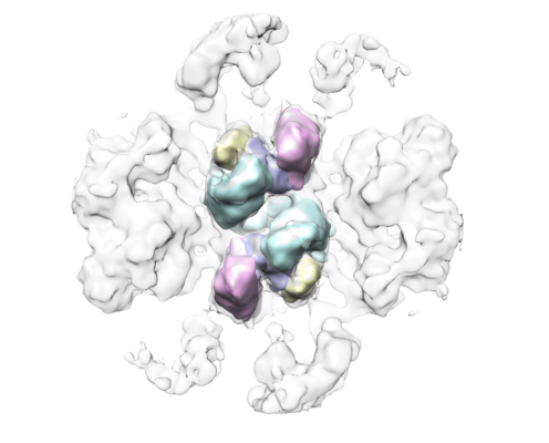 emd_0540.png
 http://ftp.pdbj.org/pub/emdb/structures/EMD-0540


 Z (Sec.)
 Y (Row.)
 X (Col.)


 Sample components
 Processing
 Electron microscopy
 FIELD EMISSION GUN