 Movie
Movie Controller

[English] 日本語
 Yorodumi
- PDB-6w51: Structure of the antibody fragment H2 in complex with HLA-A*02:01... -
+ Open data

- Basic information
| Entry | Database: PDB / ID: 6w51 | ||||||
|---|---|---|---|---|---|---|---|
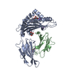
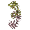
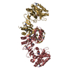
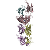
| Title | Structure of the antibody fragment H2 in complex with HLA-A*02:01/p53R175H | ||||||
 Components Components |
| ||||||
 Keywords Keywords | IMMUNE SYSTEM / Immunotherapy / MHC-I / HLA-A2 / p53 | ||||||
| Function / homology |  Function and homology information Function and homology informationnegative regulation of helicase activity / Loss of function of TP53 in cancer due to loss of tetramerization ability / Regulation of TP53 Expression / signal transduction by p53 class mediator / negative regulation of G1 to G0 transition / negative regulation of glucose catabolic process to lactate via pyruvate / Transcriptional activation of cell cycle inhibitor p21 / regulation of intrinsic apoptotic signaling pathway by p53 class mediator / negative regulation of pentose-phosphate shunt / Activation of NOXA and translocation to mitochondria ...negative regulation of helicase activity / Loss of function of TP53 in cancer due to loss of tetramerization ability / Regulation of TP53 Expression / signal transduction by p53 class mediator / negative regulation of G1 to G0 transition / negative regulation of glucose catabolic process to lactate via pyruvate / Transcriptional activation of cell cycle inhibitor p21 / regulation of intrinsic apoptotic signaling pathway by p53 class mediator / negative regulation of pentose-phosphate shunt / Activation of NOXA and translocation to mitochondria / ATP-dependent DNA/DNA annealing activity / regulation of cell cycle G2/M phase transition / oligodendrocyte apoptotic process / negative regulation of miRNA processing / intrinsic apoptotic signaling pathway in response to hypoxia / oxidative stress-induced premature senescence / regulation of tissue remodeling / positive regulation of thymocyte apoptotic process / positive regulation of mitochondrial membrane permeability / germ cell nucleus / regulation of fibroblast apoptotic process / bone marrow development / circadian behavior / cellular response to actinomycin D / histone deacetylase regulator activity / positive regulation of programmed necrotic cell death / : / regulation of mitochondrial membrane permeability involved in apoptotic process / RUNX3 regulates CDKN1A transcription / T cell proliferation involved in immune response / TP53 Regulates Transcription of Death Receptors and Ligands / Activation of PUMA and translocation to mitochondria / TP53 regulates transcription of additional cell cycle genes whose exact role in the p53 pathway remain uncertain / mRNA transcription / negative regulation of glial cell proliferation / regulation of DNA damage response, signal transduction by p53 class mediator / Regulation of TP53 Activity through Association with Co-factors / negative regulation of neuroblast proliferation / Formation of Senescence-Associated Heterochromatin Foci (SAHF) / mitochondrial DNA repair / T cell lineage commitment / thymocyte apoptotic process / ER overload response / TP53 Regulates Transcription of Caspase Activators and Caspases / cardiac septum morphogenesis / B cell lineage commitment / entrainment of circadian clock by photoperiod / negative regulation of DNA replication / Zygotic genome activation (ZGA) / negative regulation of mitophagy / TP53 Regulates Transcription of Genes Involved in Cytochrome C Release / necroptotic process / PI5P Regulates TP53 Acetylation / Association of TriC/CCT with target proteins during biosynthesis / positive regulation of release of cytochrome c from mitochondria / negative regulation of telomere maintenance via telomerase / SUMOylation of transcription factors / TP53 regulates transcription of several additional cell death genes whose specific roles in p53-dependent apoptosis remain uncertain / rRNA transcription / negative regulation of reactive oxygen species metabolic process / TFIID-class transcription factor complex binding / intrinsic apoptotic signaling pathway by p53 class mediator / Transcriptional Regulation by VENTX / cellular response to UV-C / viral process / neuroblast proliferation / intrinsic apoptotic signaling pathway in response to endoplasmic reticulum stress / replicative senescence / Pyroptosis / positive regulation of RNA polymerase II transcription preinitiation complex assembly / intrinsic apoptotic signaling pathway in response to DNA damage by p53 class mediator / general transcription initiation factor binding / chromosome organization / positive regulation of execution phase of apoptosis / hematopoietic stem cell differentiation / type II interferon-mediated signaling pathway / embryonic organ development / TP53 Regulates Transcription of Genes Involved in G1 Cell Cycle Arrest / response to X-ray / antigen processing and presentation of peptide antigen via MHC class I / hematopoietic progenitor cell differentiation / somitogenesis / negative regulation of stem cell proliferation / core promoter sequence-specific DNA binding / glial cell proliferation / cellular response to glucose starvation / negative regulation of fibroblast proliferation / cis-regulatory region sequence-specific DNA binding / mitophagy / Regulation of TP53 Activity through Acetylation / negative regulation of proteolysis / mitotic G1 DNA damage checkpoint signaling / positive regulation of intrinsic apoptotic signaling pathway / cardiac muscle cell apoptotic process / response to salt stress / transcription repressor complex / 14-3-3 protein binding / gastrulation / positive regulation of cardiac muscle cell apoptotic process / transforming growth factor beta receptor signaling pathway Similarity search - Function | ||||||
| Biological species |  Homo sapiens (human) Homo sapiens (human) | ||||||
| Method |  X-RAY DIFFRACTION / SYNCHROTRON / MOLECULAR REPLACEMENT / Resolution: 3.53 Å X-RAY DIFFRACTION / SYNCHROTRON / MOLECULAR REPLACEMENT / Resolution: 3.53 Å | ||||||
 Authors Authors | Wright, K.M. / Gabelli, S.B. | ||||||
 Citation Citation | Journal: Science / Year: 2021 Title: Targeting a neoantigen derived from a common TP53 mutation. Authors: Hsiue, E.H. / Wright, K.M. / Douglass, J. / Hwang, M.S. / Mog, B.J. / Pearlman, A.H. / Paul, S. / DiNapoli, S.R. / Konig, M.F. / Wang, Q. / Schaefer, A. / Miller, M.S. / Skora, A.D. / ...Authors: Hsiue, E.H. / Wright, K.M. / Douglass, J. / Hwang, M.S. / Mog, B.J. / Pearlman, A.H. / Paul, S. / DiNapoli, S.R. / Konig, M.F. / Wang, Q. / Schaefer, A. / Miller, M.S. / Skora, A.D. / Azurmendi, P.A. / Murphy, M.B. / Liu, Q. / Watson, E. / Li, Y. / Pardoll, D.M. / Bettegowda, C. / Papadopoulos, N. / Kinzler, K.W. / Vogelstein, B. / Gabelli, S.B. / Zhou, S. | ||||||
| History |
|
- Structure visualization
| Structure viewer | Molecule: MolmilJmol/JSmol |
|---|
- Downloads & links
-Download
| PDBx/mmCIF format | 6w51.cif.gz | 638.8 KB | Display | PDBx/mmCIF format |
|---|---|---|---|---|
| PDB format | pdb6w51.ent.gz | 527.2 KB | Display | PDB format |
| PDBx/mmJSON format | 6w51.json.gz | Tree view | PDBx/mmJSON format | |
| Others |  Other downloads Other downloads |
-Validation report
| Arichive directory | https://data.pdbj.org/pub/pdb/validation_reports/w5/6w51ftp://data.pdbj.org/pub/pdb/validation_reports/w5/6w51 | HTTPS FTP |
|---|
-Related structure data


-Links
 PDBj
PDBj


- Assembly
| Deposited unit | 
| ||||||||
|---|---|---|---|---|---|---|---|---|---|
| 1 | 
| ||||||||
| 2 | 
| ||||||||
| 3 | 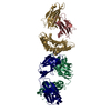
| ||||||||
| 4 | 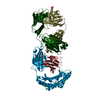
| ||||||||
| Unit cell |
|
-Components
-Protein , 2 types, 8 molecules ADGJBEHK
| #1: Protein | Mass: 34183.738 Da / Num. of mol.: 4 Source method: isolated from a genetically manipulated source Source: (gene. exp.) Homo sapiens (human) / Gene: HLA-A / Plasmid: pET3a / Production host:  #2: Protein | Mass: 13732.547 Da / Num. of mol.: 4 Source method: isolated from a genetically manipulated source Source: (gene. exp.) Homo sapiens (human) / Gene: B2M, CDABP0092, HDCMA22P / Plasmid: pET3a / Production host: |
|---|
-Protein/peptide , 1 types, 4 molecules CFIL
| #3: Protein/peptide | Mass: 1114.320 Da / Num. of mol.: 4 / Mutation: R175H / Source method: obtained synthetically / Source: (synth.) Homo sapiens (human) / References: UniProt: P04637 |
|---|
-Antibody , 2 types, 8 molecules MOQSNPRT
| #4: Antibody | Mass: 23780.512 Da / Num. of mol.: 4 Source method: isolated from a genetically manipulated source Source: (gene. exp.) Homo sapiens (human) / Plasmid: pcDNA3.1 / Cell line (production host): HEK293F / Production host: Homo sapiens (human)#5: Antibody | Mass: 23698.314 Da / Num. of mol.: 4 Source method: isolated from a genetically manipulated source Source: (gene. exp.) Homo sapiens (human) / Plasmid: pcDNA3.1 / Cell line (production host): HEK293F / Production host: Homo sapiens (human) |
|---|
-Non-polymers , 2 types, 2 molecules 

| #6: Chemical | ChemComp-PO4 /  Mass: 94.971 Da / Num. of mol.: 1 / Source method: obtained synthetically / Formula: PO4 Mass: 94.971 Da / Num. of mol.: 1 / Source method: obtained synthetically / Formula: PO4 |
|---|---|
| #7: Water | ChemComp-HOH / Mass: 18.015 Da / Num. of mol.: 1 / Source method: isolated from a natural source / Formula: H2O |
-Details
| Has ligand of interest | N |
|---|---|
| Has protein modification | Y |
-Experimental details
-Experiment
| Experiment | Method: X-RAY DIFFRACTION / Number of used crystals: 1 |
|---|
- Sample preparation
| Crystal | Density Matthews: 2.46 Å3/Da / Density % sol: 49.95 % / Mosaicity: 0.18 ° |
|---|---|
| Crystal grow | Temperature: 277 K / Method: vapor diffusion / Details: 0.2 M Ammonium chloride, 20% w/v PEG 3350 |
-Data collection
| Diffraction | Mean temperature: 100 K / Serial crystal experiment: N | ||||||||||||||||||||||||||||||
|---|---|---|---|---|---|---|---|---|---|---|---|---|---|---|---|---|---|---|---|---|---|---|---|---|---|---|---|---|---|---|---|
| Diffraction source | Source: SYNCHROTRON / Site: NSLS-II  / Beamline: 17-ID-2 / Wavelength: 0.97931 Å / Beamline: 17-ID-2 / Wavelength: 0.97931 Å | ||||||||||||||||||||||||||||||
| Detector | Type: DECTRIS EIGER X 16M / Detector: PIXEL / Date: Feb 14, 2020 | ||||||||||||||||||||||||||||||
| Radiation | Protocol: SINGLE WAVELENGTH / Monochromatic (M) / Laue (L): M / Scattering type: x-ray | ||||||||||||||||||||||||||||||
| Radiation wavelength | Wavelength: 0.97931 Å / Relative weight: 1 | ||||||||||||||||||||||||||||||
| Reflection | Resolution: 3.53→30.37 Å / Num. obs: 43734 / % possible obs: 95.3 % / Redundancy: 2.4 % / CC1/2: 0.938 / Rmerge(I) obs: 0.247 / Rpim(I) all: 0.186 / Rrim(I) all: 0.311 / Net I/σ(I): 3.6 | ||||||||||||||||||||||||||||||
| Reflection shell | Diffraction-ID: 1
|
- Processing
| Software |
| ||||||||||||||||||||||||||||||||||||||||||||||||||||||||||||
|---|---|---|---|---|---|---|---|---|---|---|---|---|---|---|---|---|---|---|---|---|---|---|---|---|---|---|---|---|---|---|---|---|---|---|---|---|---|---|---|---|---|---|---|---|---|---|---|---|---|---|---|---|---|---|---|---|---|---|---|---|---|
| Refinement | Method to determine structure: MOLECULAR REPLACEMENT Starting model: 6O4Y, 6UJ9 Resolution: 3.53→30.37 Å / Cor.coef. Fo:Fc: 0.911 / Cor.coef. Fo:Fc free: 0.823 / SU B: 43.916 / SU ML: 0.656 / Cross valid method: THROUGHOUT / σ(F): 0 / ESU R Free: 0.816 / Stereochemistry target values: MAXIMUM LIKELIHOOD Details: HYDROGENS HAVE BEEN ADDED IN THE RIDING POSITIONS U VALUES : REFINED INDIVIDUALLY
| ||||||||||||||||||||||||||||||||||||||||||||||||||||||||||||
| Solvent computation | Ion probe radii: 0.8 Å / Shrinkage radii: 0.8 Å / VDW probe radii: 1.2 Å / Solvent model: MASK | ||||||||||||||||||||||||||||||||||||||||||||||||||||||||||||
| Displacement parameters | Biso max: 245.65 Å2 / Biso mean: 61.859 Å2 / Biso min: 0.5 Å2
| ||||||||||||||||||||||||||||||||||||||||||||||||||||||||||||
| Refinement step | Cycle: final / Resolution: 3.53→30.37 Å
| ||||||||||||||||||||||||||||||||||||||||||||||||||||||||||||
| Refine LS restraints |
| ||||||||||||||||||||||||||||||||||||||||||||||||||||||||||||
| LS refinement shell | Resolution: 3.53→3.618 Å / Rfactor Rfree error: 0
|
