 Movie
Movie Controller

[English] 日本語
 Yorodumi
- EMDB-0083: Cryo-EM structure of a rotated E. coli 70S ribosome in complex wi... -
+ Open data

- Basic information
| Entry |  | |||||||||||||||
|---|---|---|---|---|---|---|---|---|---|---|---|---|---|---|---|---|
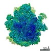
| Title | Cryo-EM structure of a rotated E. coli 70S ribosome in complex with RF3-GDPCP(RF3-only) | |||||||||||||||
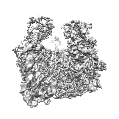
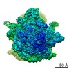
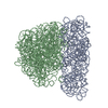
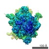
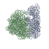
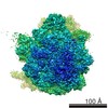
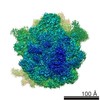
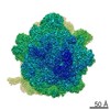
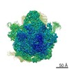
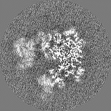
 Map data Map data | Locally filtered cryo-EM map of a rotated E. coli 70S ribosome in complex with RF3-GDPCP (RF3-only) | |||||||||||||||
 Sample Sample |
| |||||||||||||||
 Keywords Keywords | single particle cryo-EM / GTPase / ribosome / release factor RF1 / RF2 / RF3 / subunit rotation / translation termination | |||||||||||||||
| Function / homology |  Function and homology information Function and homology informationregulation of translational termination / translation release factor activity, codon specific / translation release factor activity, codon nonspecific / guanosine tetraphosphate binding / stringent response / transcription antitermination factor activity, RNA binding / ornithine decarboxylase inhibitor activity / misfolded RNA binding / Group I intron splicing / RNA folding ...regulation of translational termination / translation release factor activity, codon specific / translation release factor activity, codon nonspecific / guanosine tetraphosphate binding / stringent response / transcription antitermination factor activity, RNA binding / ornithine decarboxylase inhibitor activity / misfolded RNA binding / Group I intron splicing / RNA folding / translational termination / transcriptional attenuation / endoribonuclease inhibitor activity / positive regulation of ribosome biogenesis / RNA-binding transcription regulator activity / four-way junction DNA binding / negative regulation of cytoplasmic translation / DnaA-L2 complex / regulation of mRNA stability / translation repressor activity / negative regulation of translational initiation / negative regulation of DNA-templated DNA replication initiation / mRNA regulatory element binding translation repressor activity / positive regulation of RNA splicing / regulation of DNA-templated transcription elongation / transcription elongation factor complex / response to reactive oxygen species / cytosolic ribosome assembly / ribosome assembly / assembly of large subunit precursor of preribosome / transcription antitermination / DNA endonuclease activity / regulation of cell growth / DNA-templated transcription termination / response to radiation / maintenance of translational fidelity / mRNA 5'-UTR binding / GDP binding / regulation of translation / large ribosomal subunit / transferase activity / ribosomal small subunit assembly / ribosome binding / ribosome biogenesis / ribosomal small subunit biogenesis / 5S rRNA binding / ribosomal large subunit assembly / small ribosomal subunit / small ribosomal subunit rRNA binding / large ribosomal subunit rRNA binding / cytosolic small ribosomal subunit / cytosolic large ribosomal subunit / cytoplasmic translation / tRNA binding / defense response to bacterium / negative regulation of translation / rRNA binding / structural constituent of ribosome / ribosome / translation / innate immune response / response to antibiotic / negative regulation of DNA-templated transcription / hydrolase activity / mRNA binding / GTPase activity / GTP binding / DNA binding / RNA binding / extracellular region / zinc ion binding / membrane / cytosol / cytoplasm Similarity search - Function | |||||||||||||||
| Biological species |   | |||||||||||||||
| Method | single particle reconstruction / cryo EM / Resolution: 4.4 Å | |||||||||||||||
 Authors Authors | Graf M / Huter P | |||||||||||||||
| Funding support |  Germany, Germany,  Czech Republic, 4 items Czech Republic, 4 items
| |||||||||||||||
 Citation Citation | Journal: Nat Commun / Year: 2018 Title: Visualization of translation termination intermediates trapped by the Apidaecin 137 peptide during RF3-mediated recycling of RF1. Authors: Michael Graf / Paul Huter / Cristina Maracci / Miroslav Peterek / Marina V Rodnina / Daniel N Wilson / Abstract: During translation termination in bacteria, the release factors RF1 and RF2 are recycled from the ribosome by RF3. While high-resolution structures of the individual termination factors on the ...During translation termination in bacteria, the release factors RF1 and RF2 are recycled from the ribosome by RF3. While high-resolution structures of the individual termination factors on the ribosome exist, direct structural insight into how RF3 mediates dissociation of the decoding RFs has been lacking. Here we have used the Apidaecin 137 peptide to trap RF1 together with RF3 on the ribosome and visualize an ensemble of termination intermediates using cryo-electron microscopy. Binding of RF3 to the ribosome induces small subunit (SSU) rotation and swivelling of the head, yielding intermediate states with shifted P-site tRNAs and RF1 conformations. RF3 does not directly eject RF1 from the ribosome, but rather induces full rotation of the SSU that indirectly dislodges RF1 from its binding site. SSU rotation is coupled to the accommodation of the GTPase domain of RF3 on the large subunit (LSU), thereby promoting GTP hydrolysis and dissociation of RF3 from the ribosome. | |||||||||||||||
| History |
|
- Structure visualization
| Structure viewer | EM map: SurfViewMolmilJmol/JSmol |
|---|---|
| Supplemental images |
- Downloads & links
-EMDB archive
| Map data | emd_0083.map.gz | 86.9 MB | EMDB map data format | |
|---|---|---|---|---|
| Header (meta data) | emd-0083-v30.xmlemd-0083.xml | 83.1 KB 83.1 KB | Display Display | EMDB header |
| Images |  emd_0083.png emd_0083.png | 82.2 KB | ||
| Filedesc metadata | emd-0083.cif.gz | 14.7 KB | ||
| Others | emd_0083_additional.map.gz | 163.2 MB | ||
| Archive directory |  http://ftp.pdbj.org/pub/emdb/structures/EMD-0083ftp://ftp.pdbj.org/pub/emdb/structures/EMD-0083 http://ftp.pdbj.org/pub/emdb/structures/EMD-0083ftp://ftp.pdbj.org/pub/emdb/structures/EMD-0083 | HTTPS FTP |
-Related structure data
| Related structure data |  6gxpMC  0076C  0080C  0081C  0082C  6gwtC  6gxmC  6gxnC  6gxoC M: atomic model generated by this map C: citing same article ( |
|---|---|
| Similar structure data |
-Links
| EMDB pages | EMDB (EBI/PDBe) / EMDataResource |
|---|---|
| Related items in Molecule of the Month |


-Map
| File | Download / File: emd_0083.map.gz / Format: CCP4 / Size: 178 MB / Type: IMAGE STORED AS FLOATING POINT NUMBER (4 BYTES) | ||||||||||||||||||||||||||||||||||||
|---|---|---|---|---|---|---|---|---|---|---|---|---|---|---|---|---|---|---|---|---|---|---|---|---|---|---|---|---|---|---|---|---|---|---|---|---|---|
| Annotation | Locally filtered cryo-EM map of a rotated E. coli 70S ribosome in complex with RF3-GDPCP (RF3-only) | ||||||||||||||||||||||||||||||||||||
| Projections & slices | Image control
Images are generated by Spider. | ||||||||||||||||||||||||||||||||||||
| Voxel size | X=Y=Z: 1.061 Å | ||||||||||||||||||||||||||||||||||||
| Density |
| ||||||||||||||||||||||||||||||||||||
| Symmetry | Space group: 1 | ||||||||||||||||||||||||||||||||||||
| Details | EMDB XML:
|
 Z (Sec.)
 Y (Row.)
 X (Col.)


-Supplemental data
-Additional map: Cryo-EM map of a rotated E. coli 70S...
| File | emd_0083_additional.map | ||||||||||||
|---|---|---|---|---|---|---|---|---|---|---|---|---|---|
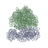
| Annotation | Cryo-EM map of a rotated E. coli 70S ribosome in complex with RF3-GDPCP (RF3-only) | ||||||||||||
| Projections & Slices |
| ||||||||||||
| Density Histograms |


- Sample components
+Entire : In-vitro reconstitution of E. coli 70S ribosomes with RF3-GDPCP, ...
+Supramolecule #1: In-vitro reconstitution of E. coli 70S ribosomes with RF3-GDPCP, ...
+Supramolecule #2: 70S ribosome
+Supramolecule #3: Apidaecin
+Macromolecule #1: 23S ribosomal RNA
+Macromolecule #2: 5S ribosomal RNA
+Macromolecule #33: 16S ribosomal RNA
+Macromolecule #3: 50S ribosomal protein L2
+Macromolecule #4: 50S ribosomal protein L3
+Macromolecule #5: 50S ribosomal protein L4
+Macromolecule #6: 50S ribosomal protein L5
+Macromolecule #7: 50S ribosomal protein L6
+Macromolecule #8: 50S ribosomal protein L9
+Macromolecule #9: 50S ribosomal protein L11
+Macromolecule #10: 50S ribosomal protein L13
+Macromolecule #11: 50S ribosomal protein L14
+Macromolecule #12: 50S ribosomal protein L15
+Macromolecule #13: 50S ribosomal protein L16
+Macromolecule #14: 50S ribosomal protein L17
+Macromolecule #15: 50S ribosomal protein L18
+Macromolecule #16: 50S ribosomal protein L19
+Macromolecule #17: 50S ribosomal protein L20
+Macromolecule #18: 50S ribosomal protein L21
+Macromolecule #19: 50S ribosomal protein L22
+Macromolecule #20: 50S ribosomal protein L23
+Macromolecule #21: 50S ribosomal protein L24
+Macromolecule #22: 50S ribosomal protein L25
+Macromolecule #23: 50S ribosomal protein L27
+Macromolecule #24: 50S ribosomal protein L28
+Macromolecule #25: 50S ribosomal protein L29
+Macromolecule #26: 50S ribosomal protein L30
+Macromolecule #27: 50S ribosomal protein L32
+Macromolecule #28: 50S ribosomal protein L33
+Macromolecule #29: 50S ribosomal protein L34
+Macromolecule #30: 50S ribosomal protein L35
+Macromolecule #31: 50S ribosomal protein L36
+Macromolecule #32: 50S ribosomal protein L10
+Macromolecule #34: 30S ribosomal protein S2
+Macromolecule #35: 30S ribosomal protein S3
+Macromolecule #36: 30S ribosomal protein S4
+Macromolecule #37: 30S ribosomal protein S5
+Macromolecule #38: 30S ribosomal protein S6
+Macromolecule #39: 30S ribosomal protein S7
+Macromolecule #40: 30S ribosomal protein S8
+Macromolecule #41: 30S ribosomal protein S9
+Macromolecule #42: 30S ribosomal protein S10
+Macromolecule #43: 30S ribosomal protein S11
+Macromolecule #44: 30S ribosomal protein S12
+Macromolecule #45: 30S ribosomal protein S13
+Macromolecule #46: 30S ribosomal protein S14
+Macromolecule #47: 30S ribosomal protein S15
+Macromolecule #48: 30S ribosomal protein S16
+Macromolecule #49: 30S ribosomal protein S17
+Macromolecule #50: 30S ribosomal protein S18
+Macromolecule #51: 30S ribosomal protein S19
+Macromolecule #52: 30S ribosomal protein S20
+Macromolecule #53: 30S ribosomal protein S21
+Macromolecule #54: Peptide chain release factor RF3
+Macromolecule #55: Apidaecin
+Macromolecule #56: PHOSPHOMETHYLPHOSPHONIC ACID GUANYLATE ESTER

-Experimental details
-Structure determination
| Method | cryo EM |
|---|---|
 Processing Processing | single particle reconstruction |
| Aggregation state | particle |
-Sample preparation
| Buffer | pH: 7.4 Component:
Details: Solutions were preapred fresh and filtered previous to usage. | |||||||||||||||
|---|---|---|---|---|---|---|---|---|---|---|---|---|---|---|---|---|
| Vitrification | Cryogen name: ETHANE / Instrument: FEI VITROBOT MARK IV |
- Electron microscopy
| Microscope | FEI TITAN KRIOS |
|---|---|
| Image recording | Film or detector model: FEI FALCON II (4k x 4k) / Detector mode: INTEGRATING / Digitization - Dimensions - Width: 4096 pixel / Digitization - Dimensions - Height: 4096 pixel / Average electron dose: 45.9 e/Å2 |
| Electron beam | Acceleration voltage: 300 kV / Electron source:  FIELD EMISSION GUN FIELD EMISSION GUN |
| Electron optics | Illumination mode: SPOT SCAN / Imaging mode: BRIGHT FIELD |
| Experimental equipment |  Model: Titan Krios / Image courtesy: FEI Company |
+Image processing
-Atomic model buiding 1
| Refinement | Space: REAL |
|---|---|
| Output model | PDB-6gxp: |
