Movie
Movie Controller
 Structure viewers
 About Yorodumi Papers


+Search query
-Structure paper
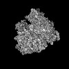
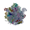
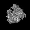
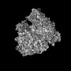
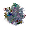
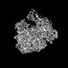
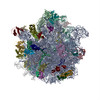
| Title | Visualization of translation termination intermediates trapped by the Apidaecin 137 peptide during RF3-mediated recycling of RF1. |
|---|---|
| Journal, issue, pages | Nat Commun, Vol. 9, Issue 1, Page 3053, Year 2018 |
| Publish date | Aug 3, 2018 |
 Authors Authors | Michael Graf / Paul Huter / Cristina Maracci / Miroslav Peterek / Marina V Rodnina / Daniel N Wilson /   |
| PubMed Abstract | During translation termination in bacteria, the release factors RF1 and RF2 are recycled from the ribosome by RF3. While high-resolution structures of the individual termination factors on the ...During translation termination in bacteria, the release factors RF1 and RF2 are recycled from the ribosome by RF3. While high-resolution structures of the individual termination factors on the ribosome exist, direct structural insight into how RF3 mediates dissociation of the decoding RFs has been lacking. Here we have used the Apidaecin 137 peptide to trap RF1 together with RF3 on the ribosome and visualize an ensemble of termination intermediates using cryo-electron microscopy. Binding of RF3 to the ribosome induces small subunit (SSU) rotation and swivelling of the head, yielding intermediate states with shifted P-site tRNAs and RF1 conformations. RF3 does not directly eject RF1 from the ribosome, but rather induces full rotation of the SSU that indirectly dislodges RF1 from its binding site. SSU rotation is coupled to the accommodation of the GTPase domain of RF3 on the large subunit (LSU), thereby promoting GTP hydrolysis and dissociation of RF3 from the ribosome. |
 External links External links | Nat Commun / PubMed:30076302 / PubMed Central |
| Methods | EM (single particle) |
| Resolution | 3.8 - 4.4 Å |
| Structure data | EMDB-0076, PDB-6gwt: EMDB-0080, PDB-6gxm: EMDB-0081, PDB-6gxn: |
| Chemicals |  ChemComp-GCP: |
| Source |
|
 Keywords Keywords | RIBOSOME / single particle cryo-EM / GTPase / release factor RF1 / RF2 / RF3 / subunit rotation / translation termination |