 Movie
Movie Controller
[English] 日本語
 Yorodumi
- SASDFN5: Wild type 2-amino-3-carboxymuconate 6-semialdehyde decarboxylase,... -
+ Open data

- Basic information
| Entry | Database: SASBDB / ID: SASDFN5 |
|---|---|
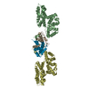
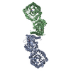
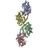
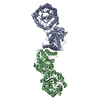
 Sample Sample | Wild type 2-amino-3-carboxymuconate 6-semialdehyde decarboxylase, ACMSD tetramer, at pH 7.0
|
| Function / homology |  Function and homology information Function and homology informationaminocarboxymuconate-semialdehyde decarboxylase / aminocarboxymuconate-semialdehyde decarboxylase activity / secondary metabolic process / hydrolase activity / metal ion binding / cytosol Similarity search - Function |
| Biological species |  Pseudomonas fluorescens (bacteria) Pseudomonas fluorescens (bacteria) |
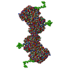
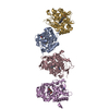
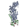
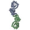
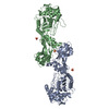
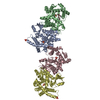
 Citation Citation | Journal: J Biol Chem / Year: 2019 Title: Quaternary structure of α-amino-β-carboxymuconate-ϵ-semialdehyde decarboxylase (ACMSD) controls its activity. Authors: Yu Yang / Ian Davis / Tsutomu Matsui / Ivan Rubalcava / Aimin Liu /  Abstract: α-Amino-β-carboxymuconate-ϵ-semialdehyde decarboxylase (ACMSD) plays an important role in l-tryptophan degradation via the kynurenine pathway. ACMSD forms a homodimer and is functionally inactive ...α-Amino-β-carboxymuconate-ϵ-semialdehyde decarboxylase (ACMSD) plays an important role in l-tryptophan degradation via the kynurenine pathway. ACMSD forms a homodimer and is functionally inactive as a monomer because its catalytic assembly requires an arginine residue from a neighboring subunit. However, how the oligomeric state and self-association of ACMSD are controlled in solution remains unexplored. Here, we demonstrate that ACMSD from can self-assemble into homodimer, tetramer, and higher-order structures. Using size-exclusion chromatography coupled with small-angle X-ray scattering (SEC-SAXS) analysis, we investigated the ACMSD tetramer structure, and fitting the SAXS data with X-ray crystal structures of the monomeric component, we could generate a pseudo-atomic structure of the tetramer. This analysis revealed a tetramer model of ACMSD as a head-on dimer of dimers. We observed that the tetramer is catalytically more active than the dimer and is in equilibrium with the monomer and dimer. Substituting a critical residue of the dimer-dimer interface, His-110, altered the tetramer dissociation profile by increasing the higher-order oligomer portion in solution without changing the X-ray crystal structure. ACMSD self-association was affected by pH, ionic strength, and other electrostatic interactions. Alignment of ACMSD sequences revealed that His-110 is highly conserved in a few bacteria that utilize nitrobenzoic acid as a sole source of carbon and energy, suggesting a dedicated functional role of ACMSD's self-assembly into the tetrameric and higher-order structures. These results indicate that the dynamic oligomerization status potentially regulates ACMSD activity and that SEC-SAXS coupled with X-ray crystallography is a powerful tool for studying protein self-association. |
 Contact author Contact author |
|
- Structure visualization
| Structure viewer | Molecule: MolmilJmol/JSmol |
|---|
- Downloads & links
-Data source
| SASBDB page |  SASDFN5 SASDFN5 |
|---|
-Related structure data


-External links
| Related items in Molecule of the Month |
|---|


-Models
| Model #2959 |  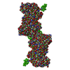 Type: mix / Radius of dummy atoms: 1.90 A / Chi-square value: 0.25 / P-value: 0.015811  Search similar-shape structures of this assembly by Omokage search (details) Search similar-shape structures of this assembly by Omokage search (details) |
|---|
-Sample
| Sample | Name: Wild type 2-amino-3-carboxymuconate 6-semialdehyde decarboxylase, ACMSD tetramer, at pH 7.0 |
|---|---|
| Buffer | Name: 25 mM HEPES, 5 mM DTT / pH: 7 |
| Entity #1627 | Name: ACMSD / Type: protein Description: 2-amino-3-carboxymuconate 6-semialdehyde decarboxylase Formula weight: 39.661 / Num. of mol.: 4 / Source: Pseudomonas fluorescens / References: UniProt: Q83V25 Sequence: MGHHHHHHHH HHSSGHIEGR HMKKPRIDMH SHFFPRISEQ EAAKFDANHA PWLQVSAKGD TGSIMMGKNN FRPVYQALWD PAFRIEEMDA QGVDVQVTCA TPVMFGYTWE ANKAAQWAER MNDFALEFAA HNPQRIKVLA QVPLQDLDLA CKEASRAVAA GHLGIQIGNH ...Sequence: MGHHHHHHHH HHSSGHIEGR HMKKPRIDMH SHFFPRISEQ EAAKFDANHA PWLQVSAKGD TGSIMMGKNN FRPVYQALWD PAFRIEEMDA QGVDVQVTCA TPVMFGYTWE ANKAAQWAER MNDFALEFAA HNPQRIKVLA QVPLQDLDLA CKEASRAVAA GHLGIQIGNH LGDKDLDDAT LEAFLTHCAN EDIPILVHPW DMMGGQRMKK WMLPWLVAMP AETQLAILSL ILSGAFERIP KSLKICFGHG GGSFAFLLGR VDNAWRHRDI VREDCPRPPS EYVDRFFVDS AVFNPGALEL LVSVMGEDRV MLGSDYPFPL GEQKIGGLVL SSNLGESAKD KIISGNASKF FNINV |
-Experimental information
| Beam | Instrument name: Stanford Synchrotron Radiation Lightsource (SSRL) BL4-2 City: Menlo Park, CA / 国: USA / Type of source: X-ray synchrotron / Wavelength: 0.1088 Å / Dist. spec. to detc.: 1.7 mm | |||||||||||||||||||||||||||||||||
|---|---|---|---|---|---|---|---|---|---|---|---|---|---|---|---|---|---|---|---|---|---|---|---|---|---|---|---|---|---|---|---|---|---|---|
| Detector | Name: Pilatus3 X 1M / Pixsize x: 0.172 mm | |||||||||||||||||||||||||||||||||
| Scan |  Measurement date: Jan 10, 2018 / Storage temperature: 25 °C / Cell temperature: 25 °C / Exposure time: 1 sec. / Number of frames: 500 / Unit: 1/A /
| |||||||||||||||||||||||||||||||||
| Distance distribution function P(R) | 
| |||||||||||||||||||||||||||||||||
| Result | 
|
