 Movie
Movie Controller

[English] 日本語
 Yorodumi
- PDB-6mgt: Crystal structure of alpha-Amino-beta-Carboxymuconate-epsilon-Sem... -
+ Open data

- Basic information
| Entry | Database: PDB / ID: 6mgt | ||||||||||||
|---|---|---|---|---|---|---|---|---|---|---|---|---|---|
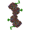
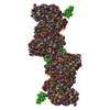
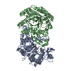
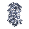
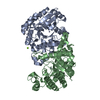
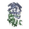
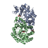
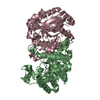
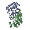
| Title | Crystal structure of alpha-Amino-beta-Carboxymuconate-epsilon-Semialdehyde Decarboxylase Mutant H110A | ||||||||||||
 Components Components | 2-amino-3-carboxymuconate 6-semialdehyde decarboxylase | ||||||||||||
 Keywords Keywords | LYASE / Holo structure / Decarboxylase | ||||||||||||
| Function / homology |  Function and homology information Function and homology informationaminocarboxymuconate-semialdehyde decarboxylase / aminocarboxymuconate-semialdehyde decarboxylase activity / secondary metabolic process / hydrolase activity / metal ion binding / cytosol Similarity search - Function | ||||||||||||
| Biological species |  Pseudomonas fluorescens (bacteria) Pseudomonas fluorescens (bacteria) | ||||||||||||
| Method |  X-RAY DIFFRACTION / SYNCHROTRON / MOLECULAR REPLACEMENT / Resolution: 2.77 Å X-RAY DIFFRACTION / SYNCHROTRON / MOLECULAR REPLACEMENT / Resolution: 2.77 Å | ||||||||||||
 Authors Authors | Yang, Y. / Daivs, I. / Matsui, T. / Rubalcava, I. / Liu, A. | ||||||||||||
| Funding support |  United States, 3items United States, 3items
| ||||||||||||
 Citation Citation | Journal: J Biol Chem / Year: 2019 Title: Quaternary structure of α-amino-β-carboxymuconate-ϵ-semialdehyde decarboxylase (ACMSD) controls its activity. Authors: Yu Yang / Ian Davis / Tsutomu Matsui / Ivan Rubalcava / Aimin Liu / Abstract: α-Amino-β-carboxymuconate-ϵ-semialdehyde decarboxylase (ACMSD) plays an important role in l-tryptophan degradation via the kynurenine pathway. ACMSD forms a homodimer and is functionally inactive ...α-Amino-β-carboxymuconate-ϵ-semialdehyde decarboxylase (ACMSD) plays an important role in l-tryptophan degradation via the kynurenine pathway. ACMSD forms a homodimer and is functionally inactive as a monomer because its catalytic assembly requires an arginine residue from a neighboring subunit. However, how the oligomeric state and self-association of ACMSD are controlled in solution remains unexplored. Here, we demonstrate that ACMSD from can self-assemble into homodimer, tetramer, and higher-order structures. Using size-exclusion chromatography coupled with small-angle X-ray scattering (SEC-SAXS) analysis, we investigated the ACMSD tetramer structure, and fitting the SAXS data with X-ray crystal structures of the monomeric component, we could generate a pseudo-atomic structure of the tetramer. This analysis revealed a tetramer model of ACMSD as a head-on dimer of dimers. We observed that the tetramer is catalytically more active than the dimer and is in equilibrium with the monomer and dimer. Substituting a critical residue of the dimer-dimer interface, His-110, altered the tetramer dissociation profile by increasing the higher-order oligomer portion in solution without changing the X-ray crystal structure. ACMSD self-association was affected by pH, ionic strength, and other electrostatic interactions. Alignment of ACMSD sequences revealed that His-110 is highly conserved in a few bacteria that utilize nitrobenzoic acid as a sole source of carbon and energy, suggesting a dedicated functional role of ACMSD's self-assembly into the tetrameric and higher-order structures. These results indicate that the dynamic oligomerization status potentially regulates ACMSD activity and that SEC-SAXS coupled with X-ray crystallography is a powerful tool for studying protein self-association. | ||||||||||||
| History |
|
- Structure visualization
| Structure viewer | Molecule: MolmilJmol/JSmol |
|---|
- Downloads & links
-Download
| PDBx/mmCIF format | 6mgt.cif.gz | 142.8 KB | Display | PDBx/mmCIF format |
|---|---|---|---|---|
| PDB format | pdb6mgt.ent.gz | 110 KB | Display | PDB format |
| PDBx/mmJSON format | 6mgt.json.gz | Tree view | PDBx/mmJSON format | |
| Others |  Other downloads Other downloads |
-Validation report
| Arichive directory | https://data.pdbj.org/pub/pdb/validation_reports/mg/6mgtftp://data.pdbj.org/pub/pdb/validation_reports/mg/6mgt | HTTPS FTP |
|---|
-Related structure data
| Related structure data |  6mgsC  2hbvS S: Starting model for refinement C: citing same article ( |
|---|---|
| Similar structure data | |
| Other databases |
|

-Links
 PDBj
PDBj


- Assembly
| Deposited unit | 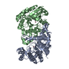
| |||||||||||||||||||||||||||||||||||||||||||||||||||||||||||||||||||||||||
|---|---|---|---|---|---|---|---|---|---|---|---|---|---|---|---|---|---|---|---|---|---|---|---|---|---|---|---|---|---|---|---|---|---|---|---|---|---|---|---|---|---|---|---|---|---|---|---|---|---|---|---|---|---|---|---|---|---|---|---|---|---|---|---|---|---|---|---|---|---|---|---|---|---|---|
| 1 |
| |||||||||||||||||||||||||||||||||||||||||||||||||||||||||||||||||||||||||
| Unit cell |
| |||||||||||||||||||||||||||||||||||||||||||||||||||||||||||||||||||||||||
| Noncrystallographic symmetry (NCS) | NCS domain:
NCS domain segments:
|