 Movie
Movie Controller

[English] 日本語
 Yorodumi
- PDB-4era: Evidence for a Dual Role of an Active Site Histidine in alpha-Ami... -
+ Open data

- Basic information
| Entry | Database: PDB / ID: 4era | ||||||
|---|---|---|---|---|---|---|---|
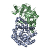
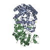
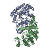
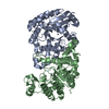
| Title | Evidence for a Dual Role of an Active Site Histidine in alpha-Amino-beta-Carboxymuconate-epsilon-Semialdehyde Decarboxylase | ||||||
 Components Components | 2-amino-3-carboxymuconate 6-semialdehyde decarboxylase | ||||||
 Keywords Keywords | LYASE / tim-barrel / decarboxylase / Metal-binding / Co(II) | ||||||
| Function / homology |  Function and homology information Function and homology informationaminocarboxymuconate-semialdehyde decarboxylase / aminocarboxymuconate-semialdehyde decarboxylase activity / secondary metabolic process / hydrolase activity / metal ion binding / cytosol Similarity search - Function | ||||||
| Biological species |  Pseudomonas fluorescens (bacteria) Pseudomonas fluorescens (bacteria) | ||||||
| Method |  X-RAY DIFFRACTION / SYNCHROTRON / MOLECULAR REPLACEMENT / Resolution: 2.398 Å X-RAY DIFFRACTION / SYNCHROTRON / MOLECULAR REPLACEMENT / Resolution: 2.398 Å | ||||||
 Authors Authors | Huo, L. / Fielding, A.J. / Chen, Y. / Li, T. / Iwaki, H. / Hosler, J.P. / Chen, L. / Hasegawa, Y. / Que Jr., L. / Liu, A. | ||||||
 Citation Citation | Journal: Biochemistry / Year: 2012 Title: Evidence for a Dual Role of an Active Site Histidine in alpha-Amino-beta-Carboxymuconate-epsilon-Semialdehyde Decarboxylase Authors: Huo, L. / Fielding, A.J. / Chen, Y. / Li, T. / Iwaki, H. / Hosler, J.P. / Chen, L. / Hasegawa, Y. / Que, L. / Liu, A. | ||||||
| History |
|
- Structure visualization
| Structure viewer | Molecule: MolmilJmol/JSmol |
|---|
- Downloads & links
-Download
| PDBx/mmCIF format | 4era.cif.gz | 140.9 KB | Display | PDBx/mmCIF format |
|---|---|---|---|---|
| PDB format | pdb4era.ent.gz | 110.8 KB | Display | PDB format |
| PDBx/mmJSON format | 4era.json.gz | Tree view | PDBx/mmJSON format | |
| Others |  Other downloads Other downloads |
-Validation report
| Arichive directory | https://data.pdbj.org/pub/pdb/validation_reports/er/4eraftp://data.pdbj.org/pub/pdb/validation_reports/er/4era | HTTPS FTP |
|---|
-Related structure data
| Related structure data |  4epkC  4ergC  4eriC  2hbxS C: citing same article ( S: Starting model for refinement |
|---|---|
| Similar structure data |


-Links
 PDBj
PDBj


- Assembly
| Deposited unit | 
| ||||||||
|---|---|---|---|---|---|---|---|---|---|
| 1 |
| ||||||||
| Unit cell |
| ||||||||
| Details | THE BIOLOGICAL UNIT IS A MONOMER. THERE ARE 2 BIOLOGICAL UNITS IN THE ASYMMETRIC UNIT (CHAIN A AND CHAIN B). |
-Components
| #1: Protein | Mass: 37208.621 Da / Num. of mol.: 2 / Mutation: H228Y Source method: isolated from a genetically manipulated source Source: (gene. exp.) Pseudomonas fluorescens (bacteria) / Strain: BL21(DE3) / Gene: nbaD / Plasmid: pET16b / Production host: #2: Chemical |   Mass: 58.933 Da / Num. of mol.: 2 / Source method: obtained synthetically / Formula: Co Mass: 58.933 Da / Num. of mol.: 2 / Source method: obtained synthetically / Formula: Co#3: Water | ChemComp-HOH / |  Mass: 18.015 Da / Num. of mol.: 89 / Source method: isolated from a natural source / Formula: H2O Mass: 18.015 Da / Num. of mol.: 89 / Source method: isolated from a natural source / Formula: H2O |
|---|
-Experimental details
-Experiment
| Experiment | Method: X-RAY DIFFRACTION / Number of used crystals: 1 |
|---|
- Sample preparation
| Crystal | Density Matthews: 2.28 Å3/Da / Density % sol: 46.01 % |
|---|---|
| Crystal grow | Temperature: 298 K / Method: vapor diffusion, hanging drop / pH: 8.75 Details: 15% PEG 5000, 0.1M Tris, 0.2M MGCL2, pH 8.75, VAPOR DIFFUSION, HANGING DROP, temperature 298K |
-Data collection
| Diffraction | Mean temperature: 100 K |
|---|---|
| Diffraction source | Source: SYNCHROTRON / Site: APS  / Beamline: 22-BM / Wavelength: 1 Å / Beamline: 22-BM / Wavelength: 1 Å |
| Detector | Type: MARMOSAIC 225 mm CCD / Detector: CCD / Date: Feb 24, 2012 |
| Radiation | Protocol: SINGLE WAVELENGTH / Monochromatic (M) / Laue (L): M / Scattering type: x-ray |
| Radiation wavelength | Wavelength: 1 Å / Relative weight: 1 |
| Reflection | Resolution: 2.398→83.66 Å / Num. all: 26155 / Num. obs: 26385 / % possible obs: 99.13 % / Observed criterion σ(F): 0 / Observed criterion σ(I): 0 / Redundancy: 13.9 % / Rmerge(I) obs: 0.101 / Net I/σ(I): 40.5 |
| Reflection shell | Resolution: 2.398→2.44 Å / Redundancy: 9.6 % / Rmerge(I) obs: 0.61 / Mean I/σ(I) obs: 2.89 / Num. unique all: 1294 / % possible all: 94.1 |
- Processing
| Software |
| |||||||||||||||||||||||||||||||||||||||||||||
|---|---|---|---|---|---|---|---|---|---|---|---|---|---|---|---|---|---|---|---|---|---|---|---|---|---|---|---|---|---|---|---|---|---|---|---|---|---|---|---|---|---|---|---|---|---|---|
| Refinement | Method to determine structure: MOLECULAR REPLACEMENT Starting model: PDB ENTRY 2HBX Resolution: 2.398→83.66 Å / Cor.coef. Fo:Fc: 0.951 / Cor.coef. Fo:Fc free: 0.906 / SU B: 13.048 / SU ML: 0.291 / Cross valid method: THROUGHOUT / σ(F): 1 / ESU R: 0.54 / ESU R Free: 0.325 / Stereochemistry target values: MAXIMUM LIKELIHOOD / Details: HYDROGENS HAVE BEEN USED IF PRESENT IN THE INPUT
| |||||||||||||||||||||||||||||||||||||||||||||
| Solvent computation | Ion probe radii: 0.8 Å / Shrinkage radii: 0.8 Å / VDW probe radii: 1.2 Å / Solvent model: MASK | |||||||||||||||||||||||||||||||||||||||||||||
| Displacement parameters | Biso mean: 59.574 Å2
| |||||||||||||||||||||||||||||||||||||||||||||
| Refinement step | Cycle: LAST / Resolution: 2.398→83.66 Å
| |||||||||||||||||||||||||||||||||||||||||||||
| Refine LS restraints |
| |||||||||||||||||||||||||||||||||||||||||||||
| LS refinement shell | Resolution: 2.398→2.46 Å / Total num. of bins used: 20
|
