 Movie
Movie Controller

[English] 日本語
 Yorodumi
- PDB-1q7m: Cobalamin-dependent methionine synthase (MetH) from Thermotoga ma... -
+ Open data

- Basic information
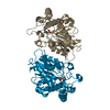
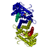
| Entry | Database: PDB / ID: 1q7m | ||||||
|---|---|---|---|---|---|---|---|
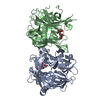
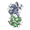
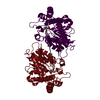
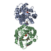
| Title | Cobalamin-dependent methionine synthase (MetH) from Thermotoga maritima (Oxidized, Monoclinic) | ||||||
 Components Components | 5-methyltetrahydrofolate S-homocysteine methyltransferase | ||||||
 Keywords Keywords | TRANSFERASE / homocysteine / methionine / folate / cobalamin / vitamin b12 | ||||||
| Function / homology |  Function and homology information Function and homology informationmethionine synthase / methionine synthase activity / homocysteine metabolic process / tetrahydrofolate metabolic process / cobalamin binding / : / methylation / metal ion binding / cytosol Similarity search - Function | ||||||
| Biological species |   Thermotoga maritima (bacteria) Thermotoga maritima (bacteria) | ||||||
| Method |  X-RAY DIFFRACTION / SYNCHROTRON / MOLECULAR REPLACEMENT / Resolution: 2.1 Å X-RAY DIFFRACTION / SYNCHROTRON / MOLECULAR REPLACEMENT / Resolution: 2.1 Å | ||||||
 Authors Authors | Evans, J.C. / Huddler, D.P. / Hilgers, M.T. / Romanchuk, G. / Matthews, R.G. / Ludwig, M.L. | ||||||
 Citation Citation | Journal: Proc.Natl.Acad.Sci.Usa / Year: 2004 Title: Structures of the N-terminal modules imply large domain motions during catalysis by methionine synthase. Authors: Evans, J.C. / Huddler, D.P. / Hilgers, M.T. / Romanchuk, G. / Matthews, R.G. / Ludwig, M.L. | ||||||
| History |
|
- Structure visualization
| Structure viewer | Molecule: MolmilJmol/JSmol |
|---|
- Downloads & links
-Download
| PDBx/mmCIF format | 1q7m.cif.gz | 224.1 KB | Display | PDBx/mmCIF format |
|---|---|---|---|---|
| PDB format | pdb1q7m.ent.gz | 180.7 KB | Display | PDB format |
| PDBx/mmJSON format | 1q7m.json.gz | Tree view | PDBx/mmJSON format | |
| Others |  Other downloads Other downloads |
-Validation report
| Arichive directory | https://data.pdbj.org/pub/pdb/validation_reports/q7/1q7mftp://data.pdbj.org/pub/pdb/validation_reports/q7/1q7m | HTTPS FTP |
|---|
-Related structure data


-Links
 PDBj
PDBj- Assembly
| Deposited unit | 
| ||||||||
|---|---|---|---|---|---|---|---|---|---|
| 1 | 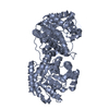
| ||||||||
| 2 | 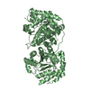
| ||||||||
| Unit cell |
|
-Components
| #1: Protein | Mass: 63633.148 Da / Num. of mol.: 2 / Fragment: MetH_Tm (residues 1-566) Source method: isolated from a genetically manipulated source Source: (gene. exp.) Thermotoga maritima (bacteria) / Plasmid: pET29a / Species (production host): Escherichia coli / Production host: #2: Water | ChemComp-HOH / |  Mass: 18.015 Da / Num. of mol.: 205 / Source method: isolated from a natural source / Formula: H2O Mass: 18.015 Da / Num. of mol.: 205 / Source method: isolated from a natural source / Formula: H2OHas protein modification | Y | |
|---|
-Experimental details
-Experiment
| Experiment | Method: X-RAY DIFFRACTION / Number of used crystals: 1 |
|---|
- Sample preparation
| Crystal | Density Matthews: 2.44 Å3/Da / Density % sol: 49.68 % | |||||||||||||||||||||||||||||||||||||||||||||||||
|---|---|---|---|---|---|---|---|---|---|---|---|---|---|---|---|---|---|---|---|---|---|---|---|---|---|---|---|---|---|---|---|---|---|---|---|---|---|---|---|---|---|---|---|---|---|---|---|---|---|---|
| Crystal grow | Temperature: 277 K / Method: vapor diffusion, sitting drop / pH: 5.2 Details: propylene glycol, glycerol, phosphate, citrate, PEG 8000, pH 5.2, VAPOR DIFFUSION, SITTING DROP, temperature 277K | |||||||||||||||||||||||||||||||||||||||||||||||||
| Crystal grow | *PLUS Temperature: 4 ℃ / pH: 5.7 / Method: vapor diffusion, sitting drop | |||||||||||||||||||||||||||||||||||||||||||||||||
| Components of the solutions | *PLUS
|
-Data collection
| Diffraction | Mean temperature: 100 K |
|---|---|
| Diffraction source | Source: SYNCHROTRON / Site: APS  / Beamline: 5ID-B / Wavelength: 1 Å / Beamline: 5ID-B / Wavelength: 1 Å |
| Detector | Type: MARRESEARCH / Detector: CCD / Date: Mar 12, 2003 |
| Radiation | Protocol: SINGLE WAVELENGTH / Monochromatic (M) / Laue (L): M / Scattering type: x-ray |
| Radiation wavelength | Wavelength: 1 Å / Relative weight: 1 |
| Reflection | Resolution: 2.1→20 Å / Num. all: 71623 / Num. obs: 58894 / % possible obs: 82.2 % / Observed criterion σ(F): 0 / Observed criterion σ(I): 0 |
| Reflection shell | Resolution: 2.1→2.2 Å / % possible all: 80.8 |
| Reflection | *PLUS Num. obs: 58895 / % possible obs: 100 % / Redundancy: 9.2 % / Rmerge(I) obs: 0.054 |
| Reflection shell | *PLUS % possible obs: 100 % / Rmerge(I) obs: 0.222 / Mean I/σ(I) obs: 8.95 |
- Processing
| Software |
| ||||||||||||||||||||
|---|---|---|---|---|---|---|---|---|---|---|---|---|---|---|---|---|---|---|---|---|---|
| Refinement | Method to determine structure: MOLECULAR REPLACEMENT / Resolution: 2.1→20 Å / σ(F): 0 / Stereochemistry target values: Engh & Huber
| ||||||||||||||||||||
| Refinement step | Cycle: LAST / Resolution: 2.1→20 Å
| ||||||||||||||||||||
| Refinement | *PLUS % reflection Rfree: 10 % / Rfactor Rfree: 0.238 / Rfactor Rwork: 0.195 | ||||||||||||||||||||
| Solvent computation | *PLUS | ||||||||||||||||||||
| Displacement parameters | *PLUS | ||||||||||||||||||||
| Refine LS restraints | *PLUS
|
