[English] 日本語
 Yorodumi
- SASDBE2: Light state solution structure of untagged Aureochrome1a- A´α-LOV-Jα -
+ Open data
- Basic information
| Entry | Database: SASBDB / ID: SASDBE2 |
|---|---|
 Sample Sample | Light state solution structure of untagged Aureochrome1a- A´α-LOV-Jα
|
| Function / homology | :  Function and homology information Function and homology information |
| Biological species |  |
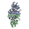
 Citation Citation |  Journal: Structure / Year: 2016 Journal: Structure / Year: 2016Title: Structure of a Native-like Aureochrome 1a LOV Domain Dimer from Phaeodactylum tricornutum. Authors: Ankan Banerjee / Elena Herman / Tilman Kottke / Lars-Oliver Essen /  Abstract: Light-oxygen-voltage (LOV) domains absorb blue light for mediating various biological responses in all three domains of life. Aureochromes from stramenopile algae represent a subfamily of ...Light-oxygen-voltage (LOV) domains absorb blue light for mediating various biological responses in all three domains of life. Aureochromes from stramenopile algae represent a subfamily of photoreceptors that differs by its inversed topology with a C-terminal LOV sensor and an N-terminal effector (basic region leucine zipper, bZIP) domain. We crystallized the LOV domain including its flanking helices, A'α and Jα, of aureochrome 1a from Phaeodactylum tricornutum in the dark state and solved the structure at 2.8 Å resolution. Both flanking helices contribute to the interface of the native-like dimer. Small-angle X-ray scattering shows light-induced conformational changes limited to the dimeric envelope as well as increased flexibility in the lit state for the flanking helices. These rearrangements are considered to be crucial for the formation of the light-activated dimer. Finally, the LOV domain of the class 2 aureochrome PtAUREO2 was shown to lack a chromophore because of steric hindrance caused by M301. |
 Contact author Contact author |
|
- Structure visualization
| Structure viewer | Molecule:  Molmil Molmil Jmol/JSmol Jmol/JSmol |
|---|
- Downloads & links
-Models
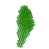
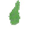
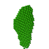
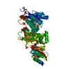
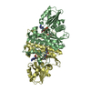
| Model #396 | 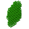 Type: dummy / Software: DAMMIN / Radius of dummy atoms: 2.75 A / Chi-square value: 0.787  Search similar-shape structures of this assembly by Omokage search (details) Search similar-shape structures of this assembly by Omokage search (details) |
|---|
- Sample
 Sample Sample | Name: Light state solution structure of untagged Aureochrome1a- A´α-LOV-Jα Specimen concentration: 2.00-8.00 |
|---|---|
| Buffer | Name: Tris / Concentration: 10.00 mM / Composition: 300 mM NaCl |
| Entity #261 | Name: PtAUREO1a-LOV / Type: protein / Description: Aureochrome1a- A´α-LOV-Jα (Light State) / Formula weight: 16.1 / Num. of mol.: 2 / Source: Phaeodactylum tricornutum / References: UniProt: JGI-49116 Sequence: GSHMDFSFIK ALQTAQQNFV VTDPSLPDNP IVYASQGFLN LTGYSLDQIL GRNCRFLQGP ETDPKAVERI RKAIEQGNDM SVCLLNYRVD GTTFWNQFFI AALRDAGGNV TNFVGVQCKV SDQYAATVTK QQEEEEEAAA NDDED |
-Experimental information
| Beam | Instrument name: ESRF BM29 / City: Grenoble / 国: France  / Type of source: X-ray synchrotron / Wavelength: 0.93 Å / Dist. spec. to detc.: 2.43 mm / Type of source: X-ray synchrotron / Wavelength: 0.93 Å / Dist. spec. to detc.: 2.43 mm | |||||||||||||||||||||
|---|---|---|---|---|---|---|---|---|---|---|---|---|---|---|---|---|---|---|---|---|---|---|
| Detector | Name: Pilatus 1M | |||||||||||||||||||||
| Scan |
| |||||||||||||||||||||
| Distance distribution function P(R) |
| |||||||||||||||||||||
| Result |
|
 Movie
Movie Controller

 SASDBE2