 Movie
Movie Controller

+ Open data

- Basic information
| Entry | Database: SASBDB / ID: SASDA58 |
|---|---|
 Sample Sample | UL26N of pseudorabies virus
|
| Function / homology |  Function and homology information Function and homology informationassemblin / nuclear capsid assembly / viral release from host cell / host cell cytoplasm / serine-type endopeptidase activity / host cell nucleus / proteolysis / identical protein binding Similarity search - Function |
| Biological species |  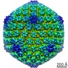 Suid herpesvirus 1 Suid herpesvirus 1 |
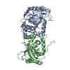
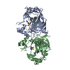
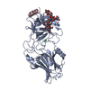
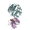
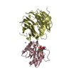
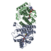
 Citation Citation | Journal: PLoS Pathog / Year: 2015 Title: Dimerization-Induced Allosteric Changes of the Oxyanion-Hole Loop Activate the Pseudorabies Virus Assemblin pUL26N, a Herpesvirus Serine Protease. Authors: Martin Zühlsdorf / Sebastiaan Werten / Barbara G Klupp / Gottfried J Palm / Thomas C Mettenleiter / Winfried Hinrichs /  Abstract: Herpesviruses encode a characteristic serine protease with a unique fold and an active site that comprises the unusual triad Ser-His-His. The protease is essential for viral replication and as such ...Herpesviruses encode a characteristic serine protease with a unique fold and an active site that comprises the unusual triad Ser-His-His. The protease is essential for viral replication and as such constitutes a promising drug target. In solution, a dynamic equilibrium exists between an inactive monomeric and an active dimeric form of the enzyme, which is believed to play a key regulatory role in the orchestration of proteolysis and capsid assembly. Currently available crystal structures of herpesvirus proteases correspond either to the dimeric state or to complexes with peptide mimetics that alter the dimerization interface. In contrast, the structure of the native monomeric state has remained elusive. Here, we present the three-dimensional structures of native monomeric, active dimeric, and diisopropyl fluorophosphate-inhibited dimeric protease derived from pseudorabies virus, an alphaherpesvirus of swine. These structures, solved by X-ray crystallography to respective resolutions of 2.05, 2.10 and 2.03 Å, allow a direct comparison of the main conformational states of the protease. In the dimeric form, a functional oxyanion hole is formed by a loop of 10 amino-acid residues encompassing two consecutive arginine residues (Arg136 and Arg137); both are strictly conserved throughout the herpesviruses. In the monomeric form, the top of the loop is shifted by approximately 11 Å, resulting in a complete disruption of the oxyanion hole and loss of activity. The dimerization-induced allosteric changes described here form the physical basis for the concentration-dependent activation of the protease, which is essential for proper virus replication. Small-angle X-ray scattering experiments confirmed a concentration-dependent equilibrium of monomeric and dimeric protease in solution. |
 Contact author Contact author |
|
- Structure visualization
| Structure viewer | Molecule: MolmilJmol/JSmol |
|---|
- Downloads & links
 SASDA58


-Models
| Model #311 |   Type: atomic / Software: Crysol / Chi-square value: 1.288  Search similar-shape structures of this assembly by Omokage search (details) Search similar-shape structures of this assembly by Omokage search (details) |
|---|---|
| Model #350 |  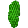 Type: dummy / Software: DAMMIN / Radius of dummy atoms: 2.25 A / Chi-square value: 0.902 Search similar-shape structures of this assembly by Omokage search (details) |
-Sample
| Sample | Name: UL26N of pseudorabies virus / Specimen concentration: 9.83 mg/ml |
|---|---|
| Buffer | Name: Tris/HCl / Concentration: 50.00 mM / pH: 7.5 Composition: 0.5 M NaCl, 0.25 M imidazole, 5% glycerol, 50 mM urea, 0.2 M MgCl2 |
| Entity #177 | Name: UL26N / Type: protein / Description: VP24 / Formula weight: 26.608 / Num. of mol.: 2 / Source: Suid herpesvirus 1 / References: UniProt: Q83417 Sequence: MGSSHHHHHH SSGLVPRGSH MGPVYVSGYL ALYDRDGGEL ALTREIVAAA LPPAGPLPIN IDHRPRCDIG AVLAVVDDDR GPFFLGVVNC PQLGAVLARA VGPDFFGDMR LSDEERLLYL LSNYLPSASL SSRRLAPGEA PDETLFAHVA LCVIGRRVGT IVVYDASPEA ...Sequence: MGSSHHHHHH SSGLVPRGSH MGPVYVSGYL ALYDRDGGEL ALTREIVAAA LPPAGPLPIN IDHRPRCDIG AVLAVVDDDR GPFFLGVVNC PQLGAVLARA VGPDFFGDMR LSDEERLLYL LSNYLPSASL SSRRLAPGEA PDETLFAHVA LCVIGRRVGT IVVYDASPEA AVAPFRQLSA RARSELLARA AESPDRERVW HMSEEALTRA LLSTAVNNML LRDRWELVAA RRREAGVRGH TYLQ |
-Experimental information
| Beam | Instrument name: PETRA III P12 / City: Hamburg / 国: Germany / Type of source: X-ray synchrotron / Wavelength: 0.12 Å / Dist. spec. to detc.: 3.1 mm | ||||||||||||||||||
|---|---|---|---|---|---|---|---|---|---|---|---|---|---|---|---|---|---|---|---|
| Detector | Name: Pilatus 2M | ||||||||||||||||||
| Scan | 
| ||||||||||||||||||
| Distance distribution function P(R) | 
| ||||||||||||||||||
| Result | 
|
