 Movie
Movie Controller


+ Open data

- Basic information
| Entry | Database: PDB / ID: 7uzm | ||||||
|---|---|---|---|---|---|---|---|
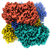
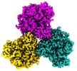
| Title | Glutamate dehydrogenase 1 from human liver | ||||||
 Components Components | Glutamate dehydrogenase 1, mitochondrial | ||||||
 Keywords Keywords | OXIDOREDUCTASE / aldehyde dehydrogenase / human liver | ||||||
| Function / homology |  Function and homology information Function and homology informationL-leucine binding / glutamate dehydrogenase [NAD(P)+] activity / tricarboxylic acid metabolic process / glutamate dehydrogenase [NAD(P)+] / glutamate biosynthetic process / glutamate dehydrogenase (NADP+) activity / glutamate dehydrogenase (NAD+) activity / Glutamate and glutamine metabolism / glutamate catabolic process / glutamine metabolic process ...L-leucine binding / glutamate dehydrogenase [NAD(P)+] activity / tricarboxylic acid metabolic process / glutamate dehydrogenase [NAD(P)+] / glutamate biosynthetic process / glutamate dehydrogenase (NADP+) activity / glutamate dehydrogenase (NAD+) activity / Glutamate and glutamine metabolism / glutamate catabolic process / glutamine metabolic process / NAD+ binding / Mitochondrial protein degradation / substantia nigra development / Transcriptional activation of mitochondrial biogenesis / positive regulation of insulin secretion / ADP binding / mitochondrial matrix / GTP binding / endoplasmic reticulum / protein homodimerization activity / mitochondrion / ATP binding / cytoplasm Similarity search - Function | ||||||
| Biological species |  Homo sapiens (human) Homo sapiens (human) | ||||||
| Method | ELECTRON MICROSCOPY / single particle reconstruction / cryo EM / Resolution: 3.24 Å | ||||||
 Authors Authors | Zhang, Z. | ||||||
| Funding support |  United States, 1items United States, 1items
| ||||||
 Citation Citation | Journal: Cell Rep / Year: 2023 Title: High-resolution structural-omics of human liver enzymes. Authors: Chih-Chia Su / Meinan Lyu / Zhemin Zhang / Masaru Miyagi / Wei Huang / Derek J Taylor / Edward W Yu / Abstract: We applied raw human liver microsome lysate to a holey carbon grid and used cryo-electron microscopy (cryo-EM) to define its composition. From this sample we identified and simultaneously determined ...We applied raw human liver microsome lysate to a holey carbon grid and used cryo-electron microscopy (cryo-EM) to define its composition. From this sample we identified and simultaneously determined high-resolution structural information for ten unique human liver enzymes involved in diverse cellular processes. Notably, we determined the structure of the endoplasmic bifunctional protein H6PD, where the N- and C-terminal domains independently possess glucose-6-phosphate dehydrogenase and 6-phosphogluconolactonase enzymatic activity, respectively. We also obtained the structure of heterodimeric human GANAB, an ER glycoprotein quality-control machinery that contains a catalytic α subunit and a noncatalytic β subunit. In addition, we observed a decameric peroxidase, PRDX4, which directly contacts a disulfide isomerase-related protein, ERp46. Structural data suggest that several glycosylations, bound endogenous compounds, and ions associate with these human liver enzymes. These results highlight the importance of cryo-EM in facilitating the elucidation of human organ proteomics at the atomic level. | ||||||
| History |
|
- Structure visualization
| Structure viewer | Molecule: MolmilJmol/JSmol |
|---|
- Downloads & links
-Download
| PDBx/mmCIF format | 7uzm.cif.gz | 490.6 KB | Display | PDBx/mmCIF format |
|---|---|---|---|---|
| PDB format | pdb7uzm.ent.gz | 407.8 KB | Display | PDB format |
| PDBx/mmJSON format | 7uzm.json.gz | Tree view | PDBx/mmJSON format | |
| Others |  Other downloads Other downloads |
-Validation report
| Summary document | 7uzm_validation.pdf.gz | 1.4 MB | Display | wwPDB validaton report |
|---|---|---|---|---|
| Full document | 7uzm_full_validation.pdf.gz | 1.4 MB | Display | |
| Data in XML | 7uzm_validation.xml.gz | 86.9 KB | Display | |
| Data in CIF | 7uzm_validation.cif.gz | 129.4 KB | Display | |
| Arichive directory | https://data.pdbj.org/pub/pdb/validation_reports/uz/7uzmftp://data.pdbj.org/pub/pdb/validation_reports/uz/7uzm | HTTPS FTP |
-Related structure data
| Related structure data | 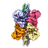 26915MC  8ekwC  8ekyC  8em2C  8emrC  8emsC 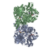 8emtC 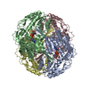 8eneC  8eojC  8eorC M: map data used to model this data C: citing same article ( |
|---|---|
| Similar structure data |


-Links
 PDBj
PDBj- Assembly
| Deposited unit | 
|
|---|---|
| 1 |
|
-Components
| #1: Protein | Mass: 61480.746 Da / Num. of mol.: 6 / Source method: isolated from a natural source / Source: (natural) Homo sapiens (human)References: UniProt: P00367, glutamate dehydrogenase [NAD(P)+] |
|---|
-Experimental details
-Experiment
| Experiment | Method: ELECTRON MICROSCOPY |
|---|---|
| EM experiment | Aggregation state: PARTICLE / 3D reconstruction method: single particle reconstruction |
- Sample preparation
| Component | Name: Glutamate dehydrogenase 1 / Type: COMPLEX / Entity ID: all / Source: NATURAL |
|---|---|
| Molecular weight | Experimental value: NO |
| Source (natural) | Organism: Homo sapiens (human) |
| Buffer solution | pH: 7.5 |
| Specimen | Embedding applied: NO / Shadowing applied: NO / Staining applied: NO / Vitrification applied: YES |
| Vitrification | Instrument: FEI VITROBOT MARK IV / Cryogen name: ETHANE |
- Electron microscopy imaging
| Experimental equipment |  Model: Titan Krios / Image courtesy: FEI Company |
|---|---|
| Microscopy | Model: FEI TITAN KRIOS |
| Electron gun | Electron source:  FIELD EMISSION GUN / Accelerating voltage: 300 kV / Illumination mode: FLOOD BEAM FIELD EMISSION GUN / Accelerating voltage: 300 kV / Illumination mode: FLOOD BEAM |
| Electron lens | Mode: BRIGHT FIELD / Nominal magnification: 82000 X / Nominal defocus max: 3291 nm / Nominal defocus min: 170 nm |
| Image recording | Electron dose: 41.25 e/Å2 / Film or detector model: GATAN K3 BIOQUANTUM (6k x 4k) |
- Processing
| Software | Name: PHENIX / Version: 1.19.2_4158: / Classification: refinement | ||||||||||||||||||||||||
|---|---|---|---|---|---|---|---|---|---|---|---|---|---|---|---|---|---|---|---|---|---|---|---|---|---|
| CTF correction | Type: PHASE FLIPPING AND AMPLITUDE CORRECTION | ||||||||||||||||||||||||
| Particle selection | Num. of particles selected: 837478 | ||||||||||||||||||||||||
| 3D reconstruction | Resolution: 3.24 Å / Resolution method: FSC 0.143 CUT-OFF / Num. of particles: 10295 / Symmetry type: POINT | ||||||||||||||||||||||||
| Refine LS restraints |
|
