 Movie
Movie Controller

[English] 日本語
 Yorodumi
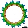
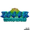
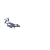
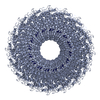
- PDB-7okn: Structure of the outer-membrane core complex (inner ring) from a ... -
+ Open data

- Basic information
| Entry | Database: PDB / ID: 7okn | ||||||
|---|---|---|---|---|---|---|---|
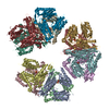
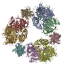
| Title | Structure of the outer-membrane core complex (inner ring) from a conjugative type IV secretion system | ||||||
 Components Components |
| ||||||
 Keywords Keywords | MEMBRANE PROTEIN / Type IV secretion system / F plasmid / outer-membrane core complex / conjugation | ||||||
| Function / homology | Type IV conjugative transfer system protein TraV / Type IV conjugative transfer system lipoprotein (TraV) / Prokaryotic membrane lipoprotein lipid attachment site profile. / Type IV conjugative transfer system lipoprotein TraV Function and homology information Function and homology information | ||||||
| Biological species |  Salmonella enterica (bacteria) Salmonella enterica (bacteria) | ||||||
| Method | ELECTRON MICROSCOPY / single particle reconstruction / cryo EM / Resolution: 3.34 Å | ||||||
 Authors Authors | Amin, H. / Ilangovan, A. / Costa, T.R.D. | ||||||
| Funding support |  United Kingdom, 1items United Kingdom, 1items
| ||||||
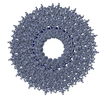
 Citation Citation | Journal: Nat Commun / Year: 2021 Title: Architecture of the outer-membrane core complex from a conjugative type IV secretion system. Authors: Himani Amin / Aravindan Ilangovan / Tiago R D Costa / Abstract: Conjugation is one of the most important processes that bacteria utilize to spread antibiotic resistance genes among bacterial populations. Interbacterial DNA transfer requires a large double ...Conjugation is one of the most important processes that bacteria utilize to spread antibiotic resistance genes among bacterial populations. Interbacterial DNA transfer requires a large double membrane-spanning nanomachine called the type 4 secretion system (T4SS) made up of the inner-membrane complex (IMC), the outer-membrane core complex (OMCC) and the conjugative pilus. The iconic F plasmid-encoded T4SS has been central in understanding conjugation for several decades, however atomic details of its structure are not known. Here, we report the structure of a complete conjugative OMCC encoded by the pED208 plasmid from E. coli, solved by cryo-electron microscopy at 3.3 Å resolution. This 2.1 MDa complex has a unique arrangement with two radial concentric rings, each having a different symmetry eventually contributing to remarkable differences in protein stoichiometry and flexibility in comparison to other OMCCs. Our structure suggests that F-OMCC is a highly dynamic complex, with implications for pilus extension and retraction during conjugation. | ||||||
| History |
|
- Structure visualization
| Movie |
Movie viewer |
|---|---|
| Structure viewer | Molecule: MolmilJmol/JSmol |
- Downloads & links
-Download
| PDBx/mmCIF format | 7okn.cif.gz | 694.1 KB | Display | PDBx/mmCIF format |
|---|---|---|---|---|
| PDB format | pdb7okn.ent.gz | 540.3 KB | Display | PDB format |
| PDBx/mmJSON format | 7okn.json.gz | Tree view | PDBx/mmJSON format | |
| Others |  Other downloads Other downloads |
-Validation report
| Arichive directory | https://data.pdbj.org/pub/pdb/validation_reports/ok/7oknftp://data.pdbj.org/pub/pdb/validation_reports/ok/7okn | HTTPS FTP |
|---|
-Related structure data
| Related structure data | 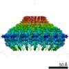 12962MC  7okoC M: map data used to model this data C: citing same article ( |
|---|---|
| Similar structure data |
-Links
 PDBj
PDBj


- Assembly
| Deposited unit | 
|
|---|---|
| 1 |
|
-Components
| #1: Protein | Mass: 48802.102 Da / Num. of mol.: 17 Source method: isolated from a genetically manipulated source Details: TraB / Source: (gene. exp.) Salmonella enterica (bacteria) / Production host: #2: Protein | Mass: 20928.650 Da / Num. of mol.: 17 Source method: isolated from a genetically manipulated source Details: TraV / Source: (gene. exp.) Salmonella enterica (bacteria) / Gene: traV, GND40_003952 / Production host: Has protein modification | Y | |
|---|
-Experimental details
-Experiment
| Experiment | Method: ELECTRON MICROSCOPY |
|---|---|
| EM experiment | Aggregation state: PARTICLE / 3D reconstruction method: single particle reconstruction |
- Sample preparation
| Component | Name: Outer-membrane core complex (inner ring) / Type: COMPLEX / Entity ID: all / Source: RECOMBINANT |
|---|---|
| Molecular weight | Experimental value: NO |
| Source (natural) | Organism: Salmonella enterica (bacteria) |
| Source (recombinant) | Organism: |
| Buffer solution | pH: 7.5 |
| Specimen | Embedding applied: NO / Shadowing applied: NO / Staining applied: NO / Vitrification applied: YES |
| Vitrification | Cryogen name: ETHANE |
- Electron microscopy imaging
| Experimental equipment |  Model: Titan Krios / Image courtesy: FEI Company |
|---|---|
| Microscopy | Model: FEI TITAN KRIOS |
| Electron gun | Electron source:  FIELD EMISSION GUN / Accelerating voltage: 300 kV / Illumination mode: SPOT SCAN FIELD EMISSION GUN / Accelerating voltage: 300 kV / Illumination mode: SPOT SCAN |
| Electron lens | Mode: BRIGHT FIELD |
| Image recording | Electron dose: 1.3 e/Å2 / Film or detector model: GATAN K3 (6k x 4k) |
- Processing
| CTF correction | Type: PHASE FLIPPING AND AMPLITUDE CORRECTION |
|---|---|
| 3D reconstruction | Resolution: 3.34 Å / Resolution method: FSC 0.143 CUT-OFF / Num. of particles: 74956 / Symmetry type: POINT |
