 ムービー
ムービー コントローラー


+ データを開く

- 基本情報
基本情報
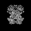
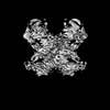
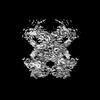
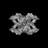
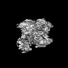
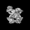
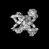
| 登録情報 | データベース: PDB / ID: 7o7p | |||||||||||||||
|---|---|---|---|---|---|---|---|---|---|---|---|---|---|---|---|---|
| タイトル | (h-alpha2M)4 activated state | |||||||||||||||
 要素 要素 | Alpha-2-macroglobulin | |||||||||||||||
 キーワード キーワード | PROTEIN BINDING / alpha2-macroglobulin / proteinase / serum proteostasis / hydrolase inhibitor | |||||||||||||||
| 機能・相同性 |  機能・相同性情報 機能・相同性情報negative regulation of complement activation, lectin pathway / interleukin-1 binding / interleukin-8 binding / tumor necrosis factor binding / HDL assembly / endopeptidase inhibitor activity / growth factor binding / Degradation of the extracellular matrix / : / platelet alpha granule lumen ...negative regulation of complement activation, lectin pathway / interleukin-1 binding / interleukin-8 binding / tumor necrosis factor binding / HDL assembly / endopeptidase inhibitor activity / growth factor binding / Degradation of the extracellular matrix / : / platelet alpha granule lumen / stem cell differentiation / serine-type endopeptidase inhibitor activity / calcium-dependent protein binding / Platelet degranulation / extracellular matrix / protease binding / blood microparticle / signaling receptor binding / enzyme binding / : / extracellular exosome / extracellular region 類似検索 - 分子機能 | |||||||||||||||
| 生物種 |  Homo sapiens (ヒト) Homo sapiens (ヒト) | |||||||||||||||
| 手法 | 電子顕微鏡法 / 単粒子再構成法 / クライオ電子顕微鏡法 / 解像度: 4.6 Å | |||||||||||||||
 データ登録者 データ登録者 | Luque, D. / Goulas, T. / Mata, C.P. / Mendes, S.R. / Gomis-Ruth, F.X. / Caston, J.R. | |||||||||||||||
| 資金援助 |  スペイン, 4件 スペイン, 4件
| |||||||||||||||
 引用 引用 | ジャーナル: Proc Natl Acad Sci U S A / 年: 2022 タイトル: Cryo-EM structures show the mechanistic basis of pan-peptidase inhibition by human α-macroglobulin. 著者: Daniel Luque / Theodoros Goulas / Carlos P Mata / Soraia R Mendes / F Xavier Gomis-Rüth / José R Castón /   要旨: Human α2-macroglobulin (hα2M) is a multidomain protein with a plethora of essential functions, including transport of signaling molecules and endopeptidase inhibition in innate immunity. Here, we ...Human α2-macroglobulin (hα2M) is a multidomain protein with a plethora of essential functions, including transport of signaling molecules and endopeptidase inhibition in innate immunity. Here, we dissected the molecular mechanism of the inhibitory function of the ∼720-kDa hα2M tetramer through eight cryo–electron microscopy (cryo-EM) structures of complexes from human plasma. In the native complex, the hα2M subunits are organized in two flexible modules in expanded conformation, which enclose a highly porous cavity in which the proteolytic activity of circulating plasma proteins is tested. Cleavage of bait regions exposed inside the cavity triggers rearrangement to a compact conformation, which closes openings and entraps the prey proteinase. After the expanded-to-compact transition, which occurs independently in the four subunits, the reactive thioester bond triggers covalent linking of the proteinase, and the receptor-binding domain is exposed on the tetramer surface for receptor-mediated clearance from circulation. These results depict the molecular mechanism of a unique suicidal inhibitory trap. | |||||||||||||||
| 履歴 |
|
- 構造の表示
構造の表示
| 構造ビューア | 分子: MolmilJmol/JSmol |
|---|
- ダウンロードとリンク
-ダウンロード
| PDBx/mmCIF形式 | 7o7p.cif.gz | 889.5 KB | 表示 | PDBx/mmCIF形式 |
|---|---|---|---|---|
| PDB形式 | pdb7o7p.ent.gz | 728 KB | 表示 | PDB形式 |
| PDBx/mmJSON形式 | 7o7p.json.gz | ツリー表示 | PDBx/mmJSON形式 | |
| その他 |  その他のダウンロード その他のダウンロード |
-検証レポート
| アーカイブディレクトリ | https://data.pdbj.org/pub/pdb/validation_reports/o7/7o7pftp://data.pdbj.org/pub/pdb/validation_reports/o7/7o7p | HTTPS FTP |
|---|
-関連構造データ
| 関連構造データ | 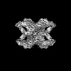 12752MC  6tavC  7o7lC  7o7mC  7o7nC  7o7oC  7o7qC  7o7rC  7o7sC M: このデータのモデリングに利用したマップデータ C: 同じ文献を引用 ( |
|---|---|
| 類似構造データ |


-リンク
 PDBj
PDBj


- 集合体
集合体
| 登録構造単位 | 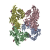
|
|---|---|
| 1 |
|
-要素
| #1: タンパク質 | 分子量: 163465.062 Da / 分子数: 4 / 由来タイプ: 天然 / 由来: (天然) Homo sapiens (ヒト) / 参照: UniProt: P01023#2: 多糖 | 2-acetamido-2-deoxy-beta-D-glucopyranose-(1-4)-2-acetamido-2-deoxy-beta-D-glucopyranose #3: 糖 | ChemComp-NAG /   タイプ: D-saccharide, beta linking / 分子量: 221.208 Da / 分子数: 20 / 由来タイプ: 合成 / 式: C8H15NO6 タイプ: D-saccharide, beta linking / 分子量: 221.208 Da / 分子数: 20 / 由来タイプ: 合成 / 式: C8H15NO6研究の焦点であるリガンドがあるか | N | Has protein modification | Y | |
|---|
-実験情報
-実験
| 実験 | 手法: 電子顕微鏡法 |
|---|---|
| EM実験 | 試料の集合状態: PARTICLE / 3次元再構成法: 単粒子再構成法 |
- 試料調製
試料調製
| 構成要素 | 名称: Human alpha-2-macroglobulin activated state / タイプ: COMPLEX / Entity ID: #1 / 由来: NATURAL |
|---|---|
| 分子量 | 値: 0.7 MDa / 実験値: NO |
| 由来(天然) | 生物種: Homo sapiens (ヒト) / 器官: Blood plasma |
| 緩衝液 | pH: 7.5 |
| 試料 | 濃度: 0.1 mg/ml / 包埋: NO / シャドウイング: NO / 染色: NO / 凍結: YES |
| 試料支持 | グリッドの材料: COPPER/RHODIUM / グリッドのサイズ: 300 divisions/in. / グリッドのタイプ: Quantifoil R2/2 |
| 急速凍結 | 装置: LEICA EM CPC / 凍結剤: ETHANE |
- 電子顕微鏡撮影
| 実験機器 |  モデル: Titan Krios / 画像提供: FEI Company |
|---|---|
| 顕微鏡 | モデル: FEI TITAN KRIOS |
| 電子銃 | 電子線源:  FIELD EMISSION GUN / 加速電圧: 300 kV / 照射モード: FLOOD BEAM FIELD EMISSION GUN / 加速電圧: 300 kV / 照射モード: FLOOD BEAM |
| 電子レンズ | モード: BRIGHT FIELD / 倍率(公称値): 47775 X / 最大 デフォーカス(公称値): 3250 nm / 最小 デフォーカス(公称値): 1000 nm |
| 試料ホルダ | 凍結剤: NITROGEN 試料ホルダーモデル: FEI TITAN KRIOS AUTOGRID HOLDER |
| 撮影 | 電子線照射量: 39.6 e/Å2 / 検出モード: COUNTING フィルム・検出器のモデル: GATAN K2 SUMMIT (4k x 4k) 実像数: 12143 |
| 画像スキャン | 動画フレーム数/画像: 32 |
- 解析
解析
| EMソフトウェア |
| ||||||||||||||||||||||||||||||||||||||||
|---|---|---|---|---|---|---|---|---|---|---|---|---|---|---|---|---|---|---|---|---|---|---|---|---|---|---|---|---|---|---|---|---|---|---|---|---|---|---|---|---|---|
| CTF補正 | タイプ: NONE | ||||||||||||||||||||||||||||||||||||||||
| 粒子像の選択 | 選択した粒子像数: 1625000 | ||||||||||||||||||||||||||||||||||||||||
| 対称性 | 点対称性: C2 (2回回転対称) | ||||||||||||||||||||||||||||||||||||||||
| 3次元再構成 | 解像度: 4.6 Å / 解像度の算出法: FSC 0.143 CUT-OFF / 粒子像の数: 118333 / アルゴリズム: FOURIER SPACE / 対称性のタイプ: POINT | ||||||||||||||||||||||||||||||||||||||||
| 原子モデル構築 | プロトコル: OTHER / 空間: REAL |
