 Movie
Movie Controller


+ Open data

- Basic information
| Entry | 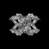 | |||||||||||||||
|---|---|---|---|---|---|---|---|---|---|---|---|---|---|---|---|---|
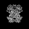
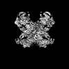
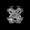
| Title | (h-alpha2M)4 trypsin-activated state | |||||||||||||||
 Map data Map data | (h-alpha2M)4 trypsin-activated state | |||||||||||||||
 Sample Sample |
| |||||||||||||||
 Keywords Keywords | alpha2-macroglobulin / proteinase / serum proteostasis / hydrolase inhibitor / PROTEIN BINDING | |||||||||||||||
| Function / homology |  Function and homology information Function and homology informationnegative regulation of complement activation, lectin pathway / interleukin-1 binding / interleukin-8 binding / tumor necrosis factor binding / HDL assembly / endopeptidase inhibitor activity / growth factor binding / Degradation of the extracellular matrix / : / platelet alpha granule lumen ...negative regulation of complement activation, lectin pathway / interleukin-1 binding / interleukin-8 binding / tumor necrosis factor binding / HDL assembly / endopeptidase inhibitor activity / growth factor binding / Degradation of the extracellular matrix / : / platelet alpha granule lumen / stem cell differentiation / serine-type endopeptidase inhibitor activity / calcium-dependent protein binding / Platelet degranulation / extracellular matrix / protease binding / blood microparticle / signaling receptor binding / enzyme binding / : / extracellular exosome / extracellular region Similarity search - Function | |||||||||||||||
| Biological species |  Homo sapiens (human) Homo sapiens (human) | |||||||||||||||
| Method | single particle reconstruction / cryo EM / Resolution: 3.6 Å | |||||||||||||||
 Authors Authors | Luque D / Goulas T | |||||||||||||||
| Funding support |  Spain, 4 items Spain, 4 items
| |||||||||||||||
 Citation Citation | Journal: Proc Natl Acad Sci U S A / Year: 2022 Title: Cryo-EM structures show the mechanistic basis of pan-peptidase inhibition by human α-macroglobulin. Authors: Daniel Luque / Theodoros Goulas / Carlos P Mata / Soraia R Mendes / F Xavier Gomis-Rüth / José R Castón /   Abstract: Human α2-macroglobulin (hα2M) is a multidomain protein with a plethora of essential functions, including transport of signaling molecules and endopeptidase inhibition in innate immunity. Here, we ...Human α2-macroglobulin (hα2M) is a multidomain protein with a plethora of essential functions, including transport of signaling molecules and endopeptidase inhibition in innate immunity. Here, we dissected the molecular mechanism of the inhibitory function of the ∼720-kDa hα2M tetramer through eight cryo–electron microscopy (cryo-EM) structures of complexes from human plasma. In the native complex, the hα2M subunits are organized in two flexible modules in expanded conformation, which enclose a highly porous cavity in which the proteolytic activity of circulating plasma proteins is tested. Cleavage of bait regions exposed inside the cavity triggers rearrangement to a compact conformation, which closes openings and entraps the prey proteinase. After the expanded-to-compact transition, which occurs independently in the four subunits, the reactive thioester bond triggers covalent linking of the proteinase, and the receptor-binding domain is exposed on the tetramer surface for receptor-mediated clearance from circulation. These results depict the molecular mechanism of a unique suicidal inhibitory trap. | |||||||||||||||
| History |
|
- Structure visualization
| Supplemental images |
|---|

- Downloads & links
-EMDB archive
| Map data | emd_12753.map.gz | 77.1 MB | EMDB map data format | |
|---|---|---|---|---|
| Header (meta data) | emd-12753-v30.xmlemd-12753.xml | 14.3 KB 14.3 KB | Display Display | EMDB header |
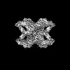
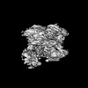
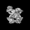
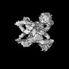
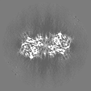
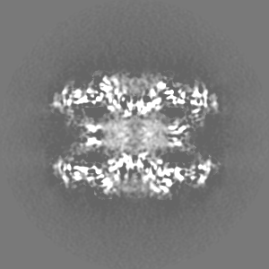
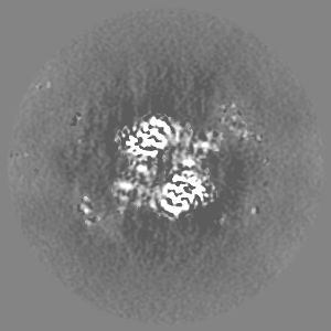
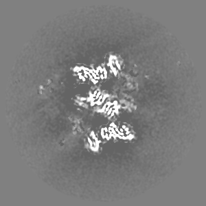
| Images |  emd_12753.png emd_12753.png | 128.4 KB | ||
| Filedesc metadata | emd-12753.cif.gz | 6.6 KB | ||
| Archive directory |  http://ftp.pdbj.org/pub/emdb/structures/EMD-12753ftp://ftp.pdbj.org/pub/emdb/structures/EMD-12753 http://ftp.pdbj.org/pub/emdb/structures/EMD-12753ftp://ftp.pdbj.org/pub/emdb/structures/EMD-12753 | HTTPS FTP |
-Related structure data
| Related structure data |  7o7qMC  6tavC  7o7lC  7o7mC  7o7nC  7o7oC  7o7pC  7o7rC  7o7sC M: atomic model generated by this map C: citing same article ( |
|---|---|
| Similar structure data |


-Links
| EMDB pages | EMDB (EBI/PDBe) / EMDataResource |
|---|---|
| Related items in Molecule of the Month |


-Map
| File | Download / File: emd_12753.map.gz / Format: CCP4 / Size: 103 MB / Type: IMAGE STORED AS FLOATING POINT NUMBER (4 BYTES) | ||||||||||||||||||||||||||||||||||||
|---|---|---|---|---|---|---|---|---|---|---|---|---|---|---|---|---|---|---|---|---|---|---|---|---|---|---|---|---|---|---|---|---|---|---|---|---|---|
| Annotation | (h-alpha2M)4 trypsin-activated state | ||||||||||||||||||||||||||||||||||||
| Projections & slices | Image control
Images are generated by Spider. | ||||||||||||||||||||||||||||||||||||
| Voxel size | X=Y=Z: 1.047 Å | ||||||||||||||||||||||||||||||||||||
| Density |
| ||||||||||||||||||||||||||||||||||||
| Symmetry | Space group: 1 | ||||||||||||||||||||||||||||||||||||
| Details | EMDB XML:
|
 Z (Sec.)
 Y (Row.)
 X (Col.)


-Supplemental data
- Sample components
-Entire : Human alpha-2-macroglobulin trypsin-activated state
| Entire | Name: Human alpha-2-macroglobulin trypsin-activated state |
|---|---|
| Components |
|
-Supramolecule #1: Human alpha-2-macroglobulin trypsin-activated state
| Supramolecule | Name: Human alpha-2-macroglobulin trypsin-activated state / type: complex / ID: 1 / Parent: 0 / Macromolecule list: #1 |
|---|---|
| Source (natural) | Organism: Homo sapiens (human) / Organ: Blood plasma |
| Molecular weight | Theoretical: 700 KDa |
-Macromolecule #1: Alpha-2-macroglobulin
| Macromolecule | Name: Alpha-2-macroglobulin / type: protein_or_peptide / ID: 1 / Number of copies: 4 / Enantiomer: LEVO |
|---|---|
| Source (natural) | Organism: Homo sapiens (human) |
| Molecular weight | Theoretical: 163.465062 KDa |
| Sequence | String: MGKNKLLHPS LVLLLLVLLP TDASVSGKPQ YMVLVPSLLH TETTEKGCVL LSYLNETVTV SASLESVRGN RSLFTDLEAE NDVLHCVAF AVPKSSSNEE VMFLTVQVKG PTQEFKKRTT VMVKNEDSLV FVQTDKSIYK PGQTVKFRVV SMDENFHPLN E LIPLVYIQ ...String: MGKNKLLHPS LVLLLLVLLP TDASVSGKPQ YMVLVPSLLH TETTEKGCVL LSYLNETVTV SASLESVRGN RSLFTDLEAE NDVLHCVAF AVPKSSSNEE VMFLTVQVKG PTQEFKKRTT VMVKNEDSLV FVQTDKSIYK PGQTVKFRVV SMDENFHPLN E LIPLVYIQ DPKGNRIAQW QSFQLEGGLK QFSFPLSSEP FQGSYKVVVQ KKSGGRTEHP FTVEEFVLPK FEVQVTVPKI IT ILEEEMN VSVCGLYTYG KPVPGHVTVS ICRKYSDASD CHGEDSQAFC EKFSGQLNSH GCFYQQVKTK VFQLKRKEYE MKL HTEAQI QEEGTVVELT GRQSSEITRT ITKLSFVKVD SHFRQGIPFF GQVRLVDGKG VPIPNKVIFI RGNEANYYSN ATTD EHGLV QFSINTTNVM GTSLTVRVNY KDRSPCYGYQ WVSEEHEEAH HTAYLVFSPS KSFVHLEPMS HELPCGHTQT VQAHY ILNG GTLLGLKKLS FYYLIMAKGG IVRTGTHGLL VKQEDMKGHF SISIPVKSDI APVARLLIYA VLPTGDVIGD SAKYDV ENC LANKVDLSFS PSQSLPASHA HLRVTAAPQS VCALRAVDQS VLLMKPDAEL SASSVYNLLP EKDLTGFPGP LNDQDNE DC INRHNVYING ITYTPVSSTN EKDMYSFLED MGLKAFTNSK IRKPKMCPQL QQYEMHGPEG LRVGFYESDV MGRGHARL V HVEEPHTETV RKYFPETWIW DLVVVNSAGV AEVGVTVPDT ITEWKAGAFC LSEDAGLGIS STASLRAFQP FFVELTMPY SVIRGEAFTL KATVLNYLPK CIRVSVQLEA SPAFLAVPVE KEQAPHCICA NGRQTVSWAV TPKSLGNVNF TVSAEALESQ ELCGTEVPS VPEHGRKDTV IKPLLVEPEG LEKETTFNSL LCPSGGEVSE ELSLKLPPNV VEESARASVS VLGDILGSAM Q NTQNLLQM PYGCGEQNMV LFAPNIYVLD YLNETQQLTP EIKSKAIGYL NTGYQRQLNY KHYDGSYSTF GERYGRNQGN TW LTAFVLK TFAQARAYIF IDEAHITQAL IWLSQRQKDN GCFRSSGSLL NNAIKGGVED EVTLSAYITI ALLEIPLTVT HPV VRNALF CLESAWKTAQ EGDHGSHVYT KALLAYAFAL AGNQDKRKEV LKSLNEEAVK KDNSVHWERP QKPKAPVGHF YEPQ APSAE VEMTSYVLLA YLTAQPAPTS EDLTSATNIV KWITKQQNAQ GGFSSTQDTV VALHALSKYG AATFTRTGKA AQVTI QSSG TFSSKFQVDN NNRLLLQQVS LPELPGEYSM KVTGEGCVYL QTSLKYNILP EKEEFPFALG VQTLPQTCDE PKAHTS FQI SLSVSYTGSR SASNMAIVDV KMVSGFIPLK PTVKMLERSN HVSRTEVSSN HVLIYLDKVS NQTLSLFFTV LQDVPVR DL KPAIVKVYDY YETDEFAIAE YNAPCSKDLG NA UniProtKB: Alpha-2-macroglobulin |
-Macromolecule #4: 2-acetamido-2-deoxy-beta-D-glucopyranose
| Macromolecule | Name: 2-acetamido-2-deoxy-beta-D-glucopyranose / type: ligand / ID: 4 / Number of copies: 20 / Formula: NAG |
|---|---|
| Molecular weight | Theoretical: 221.208 Da |
| Chemical component information |  ChemComp-NAG: |
-Experimental details
-Structure determination
| Method | cryo EM |
|---|---|
 Processing Processing | single particle reconstruction |
| Aggregation state | particle |
-Sample preparation
| Concentration | 0.1 mg/mL |
|---|---|
| Buffer | pH: 7.5 |
| Grid | Model: Quantifoil R2/2 / Material: COPPER/RHODIUM / Mesh: 300 |
| Vitrification | Cryogen name: ETHANE / Instrument: LEICA EM CPC |
- Electron microscopy
| Microscope | FEI TITAN KRIOS |
|---|---|
| Image recording | Film or detector model: GATAN K2 SUMMIT (4k x 4k) / Detector mode: COUNTING / Number real images: 6514 / Average electron dose: 40.0 e/Å2 |
| Electron beam | Acceleration voltage: 300 kV / Electron source:  FIELD EMISSION GUN FIELD EMISSION GUN |
| Electron optics | Illumination mode: FLOOD BEAM / Imaging mode: BRIGHT FIELD / Nominal defocus max: 2.5 µm / Nominal defocus min: 0.7000000000000001 µm / Nominal magnification: 47775 |
| Sample stage | Specimen holder model: FEI TITAN KRIOS AUTOGRID HOLDER / Cooling holder cryogen: NITROGEN |
| Experimental equipment |  Model: Titan Krios / Image courtesy: FEI Company |

