 Movie
Movie Controller

[English] 日本語
 Yorodumi
- PDB-7k08: Cryo-EM structure of the nonameric EscV cytosolic domain from the... -
+ Open data

- Basic information
| Entry | Database: PDB / ID: 7k08 | ||||||||||||
|---|---|---|---|---|---|---|---|---|---|---|---|---|---|
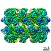
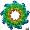
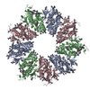
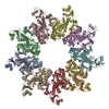
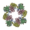
| Title | Cryo-EM structure of the nonameric EscV cytosolic domain from the type III secretion system | ||||||||||||
 Components Components | Translocator EscV | ||||||||||||
 Keywords Keywords | PROTEIN TRANSPORT / Injectisome / Nonamer / Export Apparatus / Secretion | ||||||||||||
| Function / homology |  Function and homology information Function and homology information | ||||||||||||
| Biological species |  | ||||||||||||
| Method | ELECTRON MICROSCOPY / single particle reconstruction / cryo EM / Resolution: 4.7 Å | ||||||||||||
 Authors Authors | Majewski, D.D. / Lyons, B.J.E. / Atkinson, C.E. / Strynadka, N.C.J. | ||||||||||||
| Funding support |  Canada, 3items Canada, 3items
| ||||||||||||
 Citation Citation | Journal: J Struct Biol / Year: 2020 Title: Cryo-EM analysis of the SctV cytosolic domain from the enteropathogenic E. coli T3SS injectisome. Authors: Dorothy D Majewski / Bronwyn J E Lyons / Claire E Atkinson / Natalie C J Strynadka / Abstract: The bacterial injectisome and flagella both rely on type III secretion systems for their assembly. The syringe-like injectisome creates a continuous channel between the bacterium and the host cell, ...The bacterial injectisome and flagella both rely on type III secretion systems for their assembly. The syringe-like injectisome creates a continuous channel between the bacterium and the host cell, through which signal-modulating effector proteins are secreted. The inner membrane pore protein SctV controls the hierarchy of substrate selection and may also be involved in energizing secretion. We present the 4.7 Å cryo-EM structure of the SctV cytosolic domain (SctV) from the enteropathogenic Escherichia coli injectisome. SctV forms a nonameric ring with primarily electrostatic interactions between its subunits. Molecular dynamics simulations show that monomeric SctV maintains a closed conformation, in contrast with previous studies on flagellar homologue FlhA. Comparison with substrate-bound homologues suggest that a conformational change would be required to accommodate binding partners. | ||||||||||||
| History |
|
- Structure visualization
| Movie |
Movie viewer |
|---|---|
| Structure viewer | Molecule: MolmilJmol/JSmol |
- Downloads & links
-Download
| PDBx/mmCIF format | 7k08.cif.gz | 1000.2 KB | Display | PDBx/mmCIF format |
|---|---|---|---|---|
| PDB format | pdb7k08.ent.gz | 832.9 KB | Display | PDB format |
| PDBx/mmJSON format | 7k08.json.gz | Tree view | PDBx/mmJSON format | |
| Others |  Other downloads Other downloads |
-Validation report
| Summary document | 7k08_validation.pdf.gz | 843.7 KB | Display | wwPDB validaton report |
|---|---|---|---|---|
| Full document | 7k08_full_validation.pdf.gz | 894.6 KB | Display | |
| Data in XML | 7k08_validation.xml.gz | 88.8 KB | Display | |
| Data in CIF | 7k08_validation.cif.gz | 118.1 KB | Display | |
| Arichive directory | https://data.pdbj.org/pub/pdb/validation_reports/k0/7k08ftp://data.pdbj.org/pub/pdb/validation_reports/k0/7k08 | HTTPS FTP |
-Related structure data
| Related structure data | 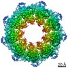 22589MC M: map data used to model this data C: citing same article ( |
|---|---|
| Similar structure data |
-Links
 PDBj
PDBj- Assembly
| Deposited unit | 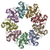
|
|---|---|
| 1 |
|
-Components
| #1: Protein | Mass: 39081.633 Da / Num. of mol.: 9 / Fragment: cytosolic domain (UNP residues 338-675) Source method: isolated from a genetically manipulated source Source: (gene. exp.) Strain: E2348/69 / EPEC / Gene: escV, E2348C_3949 / Production host: |
|---|
-Experimental details
-Experiment
| Experiment | Method: ELECTRON MICROSCOPY |
|---|---|
| EM experiment | Aggregation state: PARTICLE / 3D reconstruction method: single particle reconstruction |
- Sample preparation
| Component | Name: EscV Cytosolic Nonamer Ring / Type: COMPLEX / Entity ID: all / Source: RECOMBINANT |
|---|---|
| Molecular weight | Value: 0.35 MDa / Experimental value: NO |
| Source (natural) | Organism: |
| Source (recombinant) | Organism: |
| Buffer solution | pH: 7.5 |
| Specimen | Conc.: 1.5 mg/ml / Embedding applied: NO / Shadowing applied: NO / Staining applied: NO / Vitrification applied: YES |
| Specimen support | Grid material: COPPER / Grid mesh size: 300 divisions/in. / Grid type: Quantifoil R2/2 |
| Vitrification | Instrument: FEI VITROBOT MARK IV / Cryogen name: ETHANE / Humidity: 100 % / Chamber temperature: 277 K |
- Electron microscopy imaging
| Experimental equipment |  Model: Titan Krios / Image courtesy: FEI Company |
|---|---|
| Microscopy | Model: FEI TITAN KRIOS |
| Electron gun | Electron source:  FIELD EMISSION GUN / Accelerating voltage: 300 kV / Illumination mode: FLOOD BEAM FIELD EMISSION GUN / Accelerating voltage: 300 kV / Illumination mode: FLOOD BEAM |
| Electron lens | Mode: BRIGHT FIELD |
| Image recording | Electron dose: 60 e/Å2 / Film or detector model: FEI FALCON III (4k x 4k) |
- Processing
| EM software |
| ||||||||||||||||||
|---|---|---|---|---|---|---|---|---|---|---|---|---|---|---|---|---|---|---|---|
| CTF correction | Type: PHASE FLIPPING AND AMPLITUDE CORRECTION | ||||||||||||||||||
| Symmetry | Point symmetry: C9 (9 fold cyclic) | ||||||||||||||||||
| 3D reconstruction | Resolution: 4.7 Å / Resolution method: FSC 0.143 CUT-OFF / Num. of particles: 83566 / Symmetry type: POINT |
