Movie
Movie Controller

[English] 日本語
 Yorodumi
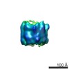
- EMDB-22590: Cryo-EM map of the stacked nonameric EscV cytosolic domain from t... -
+ Open data

- Basic information
| Entry | Database: EMDB / ID: EMD-22590 | ||||||||||||
|---|---|---|---|---|---|---|---|---|---|---|---|---|---|
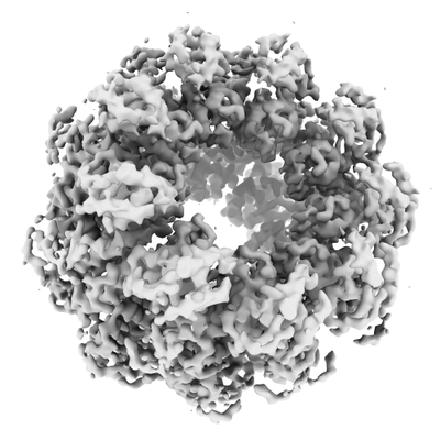
| Title | Cryo-EM map of the stacked nonameric EscV cytosolic domain from the type III secretion system | ||||||||||||
 Map data Map data | |||||||||||||
 Sample Sample |
| ||||||||||||
| Biological species |  | ||||||||||||
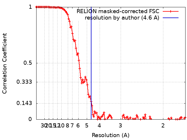
| Method | single particle reconstruction / cryo EM / Resolution: 4.6 Å | ||||||||||||
 Authors Authors | Majewski DD / Lyons BJE / Atkinson CE / Strynadka NCJ | ||||||||||||
| Funding support |  Canada, 3 items Canada, 3 items
| ||||||||||||
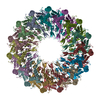
 Citation Citation | Journal: J Struct Biol / Year: 2020 Title: Cryo-EM analysis of the SctV cytosolic domain from the enteropathogenic E. coli T3SS injectisome. Authors: Dorothy D Majewski / Bronwyn J E Lyons / Claire E Atkinson / Natalie C J Strynadka / Abstract: The bacterial injectisome and flagella both rely on type III secretion systems for their assembly. The syringe-like injectisome creates a continuous channel between the bacterium and the host cell, ...The bacterial injectisome and flagella both rely on type III secretion systems for their assembly. The syringe-like injectisome creates a continuous channel between the bacterium and the host cell, through which signal-modulating effector proteins are secreted. The inner membrane pore protein SctV controls the hierarchy of substrate selection and may also be involved in energizing secretion. We present the 4.7 Å cryo-EM structure of the SctV cytosolic domain (SctV) from the enteropathogenic Escherichia coli injectisome. SctV forms a nonameric ring with primarily electrostatic interactions between its subunits. Molecular dynamics simulations show that monomeric SctV maintains a closed conformation, in contrast with previous studies on flagellar homologue FlhA. Comparison with substrate-bound homologues suggest that a conformational change would be required to accommodate binding partners. | ||||||||||||
| History |
|
- Structure visualization
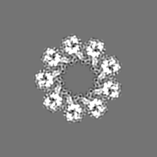
| Movie |
 Movie viewer Movie viewer |
|---|---|
| Structure viewer | EM map: SurfViewMolmilJmol/JSmol |
| Supplemental images |
- Downloads & links
-EMDB archive
| Map data | emd_22590.map.gz | 15.9 MB | EMDB map data format | |
|---|---|---|---|---|
| Header (meta data) | emd-22590-v30.xmlemd-22590.xml | 10.4 KB 10.4 KB | Display Display | EMDB header |
| FSC (resolution estimation) | emd_22590_fsc.xml | 11.5 KB | Display | FSC data file |
| Images |  emd_22590.png emd_22590.png | 86.3 KB | ||
| Archive directory |  http://ftp.pdbj.org/pub/emdb/structures/EMD-22590ftp://ftp.pdbj.org/pub/emdb/structures/EMD-22590 http://ftp.pdbj.org/pub/emdb/structures/EMD-22590ftp://ftp.pdbj.org/pub/emdb/structures/EMD-22590 | HTTPS FTP |
-Related structure data


-Links
| EMDB pages | EMDB (EBI/PDBe) / EMDataResource |
|---|
-Map
| File | Download / File: emd_22590.map.gz / Format: CCP4 / Size: 125 MB / Type: IMAGE STORED AS FLOATING POINT NUMBER (4 BYTES) | ||||||||||||||||||||||||||||||||||||||||||||||||||||||||||||||||||||
|---|---|---|---|---|---|---|---|---|---|---|---|---|---|---|---|---|---|---|---|---|---|---|---|---|---|---|---|---|---|---|---|---|---|---|---|---|---|---|---|---|---|---|---|---|---|---|---|---|---|---|---|---|---|---|---|---|---|---|---|---|---|---|---|---|---|---|---|---|---|
| Projections & slices | Image control
Images are generated by Spider. | ||||||||||||||||||||||||||||||||||||||||||||||||||||||||||||||||||||
| Voxel size | X=Y=Z: 0.852 Å | ||||||||||||||||||||||||||||||||||||||||||||||||||||||||||||||||||||
| Density |
| ||||||||||||||||||||||||||||||||||||||||||||||||||||||||||||||||||||
| Symmetry | Space group: 1 | ||||||||||||||||||||||||||||||||||||||||||||||||||||||||||||||||||||
| Details | EMDB XML:
CCP4 map header:
| ||||||||||||||||||||||||||||||||||||||||||||||||||||||||||||||||||||
 Z (Sec.)
 Y (Row.)
 X (Col.)


-Supplemental data
- Sample components
-Entire : EscV Cytosolic Nonamer Ring
| Entire | Name: EscV Cytosolic Nonamer Ring |
|---|---|
| Components |
|
-Supramolecule #1: EscV Cytosolic Nonamer Ring
| Supramolecule | Name: EscV Cytosolic Nonamer Ring / type: complex / ID: 1 / Parent: 0 / Macromolecule list: all |
|---|---|
| Source (natural) | Organism: |
| Recombinant expression | Organism: |
| Molecular weight | Theoretical: 350 KDa |
-Macromolecule #1: EscV
| Macromolecule | Name: EscV / type: protein_or_peptide / ID: 1 / Enantiomer: LEVO |
|---|---|
| Source (natural) | Organism: |
| Sequence | String: GSHMADLSNS QNISPGAEPL ILNLSSNIYS SDITQQIEVM RWNFFEESGI PLPKIIVNPV KNNDSAIEFL LYQESIYKDT LIDDTVYFEA GHAEISFEFV QEKLSTNSIV YKTNKTNQQL AHLTGMDVYA TTNDKITFLL KKLVLSNAKE FIGVQETRYL MDIMERKYNE ...String: GSHMADLSNS QNISPGAEPL ILNLSSNIYS SDITQQIEVM RWNFFEESGI PLPKIIVNPV KNNDSAIEFL LYQESIYKDT LIDDTVYFEA GHAEISFEFV QEKLSTNSIV YKTNKTNQQL AHLTGMDVYA TTNDKITFLL KKLVLSNAKE FIGVQETRYL MDIMERKYNE LVKELQRQLG LSKIVDILQR LVEENVSIRD LRTIFETLIF WSTKEKDVVI LCEYVRIALR RHILGRYSVS GTLLNVWLIG SDIENELRES IRQTSSGSYL NISPERTEQI IGFLKNIMNP TGNGVILTAL DIRRYVKKMI EGSFPSVPVL SFQEVGNNIE LKVLGTVNDF RA |
-Experimental details
-Structure determination
| Method | cryo EM |
|---|---|
 Processing Processing | single particle reconstruction |
| Aggregation state | particle |
-Sample preparation
| Concentration | 1.5 mg/mL |
|---|---|
| Buffer | pH: 7.5 |
| Vitrification | Cryogen name: ETHANE / Chamber humidity: 100 % / Chamber temperature: 277 K / Instrument: FEI VITROBOT MARK IV |
- Electron microscopy
| Microscope | FEI TITAN KRIOS |
|---|---|
| Image recording | Film or detector model: FEI FALCON III (4k x 4k) / Average electron dose: 60.0 e/Å2 |
| Electron beam | Acceleration voltage: 300 kV / Electron source:  FIELD EMISSION GUN FIELD EMISSION GUN |
| Electron optics | Illumination mode: FLOOD BEAM / Imaging mode: BRIGHT FIELD |
| Experimental equipment |  Model: Titan Krios / Image courtesy: FEI Company |