 Movie
Movie Controller

[English] 日本語
 Yorodumi
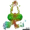
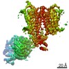
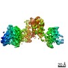
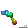
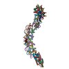
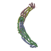
- PDB-7ewp: Cryo-EM structure of human GPR158 in complex with RGS7-Gbeta5 in ... -
+ Open data

- Basic information
| Entry | Database: PDB / ID: 7ewp | ||||||||||||
|---|---|---|---|---|---|---|---|---|---|---|---|---|---|
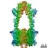
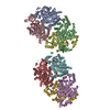
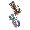
| Title | Cryo-EM structure of human GPR158 in complex with RGS7-Gbeta5 in a 2:1:1 ratio | ||||||||||||
 Components Components |
| ||||||||||||
 Keywords Keywords | SIGNALING PROTEIN / GPCR | ||||||||||||
| Function / homology |  Function and homology information Function and homology informationG protein-coupled glycine receptor activity / dendrite terminus / GTPase activator complex / dark adaptation / light adaption / G-protein gamma-subunit binding / cell tip / regulation of G protein-coupled receptor signaling pathway / G protein-coupled dopamine receptor signaling pathway / negative regulation of G protein-coupled receptor signaling pathway ...G protein-coupled glycine receptor activity / dendrite terminus / GTPase activator complex / dark adaptation / light adaption / G-protein gamma-subunit binding / cell tip / regulation of G protein-coupled receptor signaling pathway / G protein-coupled dopamine receptor signaling pathway / negative regulation of G protein-coupled receptor signaling pathway / positive regulation of neurotransmitter secretion / regulation of synapse organization / parallel fiber to Purkinje cell synapse / regulation of postsynaptic membrane potential / positive regulation of GTPase activity / G-protein alpha-subunit binding / GTPase activator activity / protein localization to plasma membrane / brain development / postsynaptic density membrane / cognition / enzyme activator activity / G beta:gamma signalling through PLC beta / Presynaptic function of Kainate receptors / Thromboxane signalling through TP receptor / Activation of G protein gated Potassium channels / Inhibition of voltage gated Ca2+ channels via Gbeta/gamma subunits / G-protein activation / Prostacyclin signalling through prostacyclin receptor / G beta:gamma signalling through CDC42 / G beta:gamma signalling through BTK / ADP signalling through P2Y purinoceptor 12 / Glucagon-type ligand receptors / transmembrane signaling receptor activity / Adrenaline,noradrenaline inhibits insulin secretion / Vasopressin regulates renal water homeostasis via Aquaporins / Glucagon-like Peptide-1 (GLP1) regulates insulin secretion / G alpha (z) signalling events / ADP signalling through P2Y purinoceptor 1 / ADORA2B mediated anti-inflammatory cytokines production / G beta:gamma signalling through PI3Kgamma / Cooperation of PDCL (PhLP1) and TRiC/CCT in G-protein beta folding / GPER1 signaling / heterotrimeric G-protein complex / G alpha (12/13) signalling events / G-protein beta-subunit binding / Inactivation, recovery and regulation of the phototransduction cascade / protein-folding chaperone binding / Thrombin signalling through proteinase activated receptors (PARs) / signaling receptor complex adaptor activity / presynaptic membrane / Ca2+ pathway / High laminar flow shear stress activates signaling by PIEZO1 and PECAM1:CDH5:KDR in endothelial cells / G alpha (i) signalling events / G alpha (s) signalling events / G alpha (q) signalling events / postsynaptic membrane / Extra-nuclear estrogen signaling / neuron projection / intracellular signal transduction / G protein-coupled receptor signaling pathway / GTPase activity / dendrite / glutamatergic synapse / signal transduction / nucleus / plasma membrane / cytoplasm / cytosol Similarity search - Function | ||||||||||||
| Biological species |  Homo sapiens (human) Homo sapiens (human) | ||||||||||||
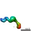
| Method | ELECTRON MICROSCOPY / single particle reconstruction / cryo EM / Resolution: 4.3 Å | ||||||||||||
 Authors Authors | Kim, Y. / Jeong, E. / Jeong, J. / Cho, Y. | ||||||||||||
| Funding support | 3items
| ||||||||||||
 Citation Citation | Journal: Nat Commun / Year: 2021 Title: Structure of the class C orphan GPCR GPR158 in complex with RGS7-Gβ5. Authors: Eunyoung Jeong / Yoojoong Kim / Jihong Jeong / Yunje Cho /  Abstract: GPR158, a class C orphan GPCR, functions in cognition, stress-induced mood control, and synaptic development. Among class C GPCRs, GPR158 is unique as it lacks a Venus flytrap-fold ligand-binding ...GPR158, a class C orphan GPCR, functions in cognition, stress-induced mood control, and synaptic development. Among class C GPCRs, GPR158 is unique as it lacks a Venus flytrap-fold ligand-binding domain and terminates Gαi/o protein signaling through the RGS7-Gβ5 heterodimer. Here, we report the cryo-EM structures of GPR158 alone and in complex with one or two RGS7-Gβ5 heterodimers. GPR158 dimerizes through Per-Arnt-Sim-fold extracellular and transmembrane (TM) domains connected by an epidermal growth factor-like linker. The TM domain (TMD) reflects both inactive and active states of other class C GPCRs: a compact intracellular TMD, conformations of the two intracellular loops (ICLs) and the TMD interface formed by TM4/5. The ICL2, ICL3, TM3, and first helix of the cytoplasmic coiled-coil provide a platform for the DHEX domain of one RGS7 and the second helix recruits another RGS7. The unique features of the RGS7-binding site underlie the selectivity of GPR158 for RGS7. | ||||||||||||
| History |
|
- Structure visualization
| Movie |
Movie viewer |
|---|---|
| Structure viewer | Molecule: MolmilJmol/JSmol |
- Downloads & links
-Download
| PDBx/mmCIF format | 7ewp.cif.gz | 565.1 KB | Display | PDBx/mmCIF format |
|---|---|---|---|---|
| PDB format | pdb7ewp.ent.gz | 427.8 KB | Display | PDB format |
| PDBx/mmJSON format | 7ewp.json.gz | Tree view | PDBx/mmJSON format | |
| Others |  Other downloads Other downloads |
-Validation report
| Arichive directory | https://data.pdbj.org/pub/pdb/validation_reports/ew/7ewpftp://data.pdbj.org/pub/pdb/validation_reports/ew/7ewp | HTTPS FTP |
|---|
-Related structure data
| Related structure data | 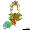 31360MC  7ewlC  7ewrC M: map data used to model this data C: citing same article ( |
|---|---|
| Similar structure data |
-Links
 PDBj
PDBj


- Assembly
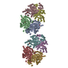
| Deposited unit | 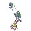
|
|---|---|
| 1 |
|
-Components
| #1: Protein | Mass: 127547.109 Da / Num. of mol.: 2 Source method: isolated from a genetically manipulated source Source: (gene. exp.) Homo sapiens (human) / Gene: GPR158 / Cell line (production host): HEK293 / Production host: Homo sapiens (human) / References: UniProt: Q5T848#2: Protein | | Mass: 61570.086 Da / Num. of mol.: 1 Source method: isolated from a genetically manipulated source Source: (gene. exp.) Homo sapiens (human) / Gene: RGS7 / Cell line (production host): HEK293 / Production host: Homo sapiens (human) / References: UniProt: P49802#3: Protein | | Mass: 43619.297 Da / Num. of mol.: 1 Source method: isolated from a genetically manipulated source Source: (gene. exp.) Homo sapiens (human) / Gene: GNB5 / Cell line (production host): HEK293 / Production host: Homo sapiens (human) / References: UniProt: O14775Has protein modification | Y | |
|---|
-Experimental details
-Experiment
| Experiment | Method: ELECTRON MICROSCOPY |
|---|---|
| EM experiment | Aggregation state: PARTICLE / 3D reconstruction method: single particle reconstruction |
- Sample preparation
| Component | Name: GPR158-RGS7-Gb5 in a 2:1:1 ratio / Type: COMPLEX / Entity ID: all / Source: RECOMBINANT |
|---|---|
| Source (natural) | Organism: Homo sapiens (human) |
| Source (recombinant) | Organism: Homo sapiens (human) |
| Buffer solution | pH: 7.5 |
| Specimen | Conc.: 4.5 mg/ml / Embedding applied: NO / Shadowing applied: NO / Staining applied: NO / Vitrification applied: YES |
| Specimen support | Grid material: COPPER / Grid mesh size: 400 divisions/in. / Grid type: C-flat-1.2/1.3 |
| Vitrification | Cryogen name: ETHANE / Humidity: 100 % |
- Electron microscopy imaging
| Experimental equipment |  Model: Talos Arctica / Image courtesy: FEI Company |
|---|---|
| Microscopy | Model: FEI TALOS ARCTICA |
| Electron gun | Electron source:  FIELD EMISSION GUN / Accelerating voltage: 200 kV / Illumination mode: OTHER FIELD EMISSION GUN / Accelerating voltage: 200 kV / Illumination mode: OTHER |
| Electron lens | Mode: DIFFRACTION |
| Image recording | Electron dose: 50 e/Å2 / Film or detector model: GATAN K3 (6k x 4k) |
- Processing
| Software | Name: PHENIX / Version: 1.14-3260_1069: / Classification: refinement |
|---|---|
| CTF correction | Type: PHASE FLIPPING AND AMPLITUDE CORRECTION |
| 3D reconstruction | Resolution: 4.3 Å / Resolution method: FSC 0.143 CUT-OFF / Num. of particles: 362999 / Symmetry type: POINT |
