 Movie
Movie Controller

[English] 日本語
 Yorodumi
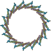
- PDB-4ckh: Helical reconstruction of ACAP1(BAR-PH domain) decorated membrane... -
+ Open data

- Basic information
| Entry | Database: PDB / ID: 4ckh | ||||||
|---|---|---|---|---|---|---|---|
| Title | Helical reconstruction of ACAP1(BAR-PH domain) decorated membrane tubules by cryo-electron microscopy | ||||||
 Components Components | ARF-GAP WITH COILED-COIL, ANK REPEAT AND PH DOMAIN-CONTAINING PROTEIN 1 | ||||||
 Keywords Keywords | SIGNALING PROTEIN / MEMBRANE REMODELING | ||||||
| Function / homology |  Function and homology information Function and homology informationGTPase activator activity / recycling endosome membrane / protein transport / zinc ion binding / membrane Similarity search - Function | ||||||
| Biological species |  HOMO SAPIENS (human) HOMO SAPIENS (human) | ||||||
| Method | ELECTRON MICROSCOPY / helical reconstruction / cryo EM / Resolution: 17 Å | ||||||
 Authors Authors | Pang, X.Y. / Fan, J. / Zhang, Y. / Zhang, K. / Gao, B.Q. / Ma, J. / Li, J. / Deng, Y.C. / Zhou, Q.J. / Hsu, V. / Sun, F. | ||||||
 Citation Citation | Journal: Dev Cell / Year: 2014 Title: A PH domain in ACAP1 possesses key features of the BAR domain in promoting membrane curvature. Authors: Xiaoyun Pang / Jun Fan / Yan Zhang / Kai Zhang / Bingquan Gao / Jun Ma / Jian Li / Yuchen Deng / Qiangjun Zhou / Edward H Egelman / Victor W Hsu / Fei Sun /   Abstract: The BAR (Bin-Amphiphysin-Rvs) domain undergoes dimerization to produce a curved protein structure, which superimposes onto membrane through electrostatic interactions to sense and impart membrane ...The BAR (Bin-Amphiphysin-Rvs) domain undergoes dimerization to produce a curved protein structure, which superimposes onto membrane through electrostatic interactions to sense and impart membrane curvature. In some cases, a BAR domain also possesses an amphipathic helix that inserts into the membrane to induce curvature. ACAP1 (Arfgap with Coil coil, Ankyrin repeat, and PH domain protein 1) contains a BAR domain. Here, we show that this BAR domain can neither bind membrane nor impart curvature, but instead requires a neighboring PH (Pleckstrin Homology) domain to achieve these functions. Specific residues within the PH domain are responsible for both membrane binding and curvature generation. The BAR domain adjacent to the PH domain instead interacts with the BAR domains of neighboring ACAP1 proteins to enable clustering at the membrane. Thus, we have uncovered the molecular basis for an unexpected and unconventional collaboration between PH and BAR domains in membrane bending. | ||||||
| History |
|
- Structure visualization
| Movie |
Movie viewer |
|---|---|
| Structure viewer | Molecule: MolmilJmol/JSmol |
- Downloads & links
-Download
| PDBx/mmCIF format | 4ckh.cif.gz | 258.3 KB | Display | PDBx/mmCIF format |
|---|---|---|---|---|
| PDB format | pdb4ckh.ent.gz | 202 KB | Display | PDB format |
| PDBx/mmJSON format | 4ckh.json.gz | Tree view | PDBx/mmJSON format | |
| Others |  Other downloads Other downloads |
-Validation report
| Arichive directory | https://data.pdbj.org/pub/pdb/validation_reports/ck/4ckhftp://data.pdbj.org/pub/pdb/validation_reports/ck/4ckh | HTTPS FTP |
|---|
-Related structure data
| Related structure data |  2547MC  2546C 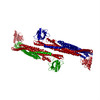 4ckgC 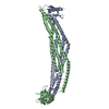 4nswC C: citing same article ( M: map data used to model this data |
|---|---|
| Similar structure data |
-Links
 PDBj
PDBj

- Assembly
| Deposited unit | 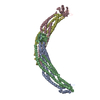
| ||||||||||||||||||||||||||||||||||||||||||||
|---|---|---|---|---|---|---|---|---|---|---|---|---|---|---|---|---|---|---|---|---|---|---|---|---|---|---|---|---|---|---|---|---|---|---|---|---|---|---|---|---|---|---|---|---|---|
| 1 | x 10
| ||||||||||||||||||||||||||||||||||||||||||||
| Noncrystallographic symmetry (NCS) | NCS oper:
|
-Components
| #1: Protein | Mass: 43334.348 Da / Num. of mol.: 4 / Fragment: BAR-PH DOMAIN, RESIDUES 1-377 Source method: isolated from a genetically manipulated source Source: (gene. exp.) HOMO SAPIENS (human) / Plasmid: PGEX-6P-1 / Production host:  Sequence details | PFAM PF03114 | |
|---|
-Experimental details
-Experiment
| Experiment | Method: ELECTRON MICROSCOPY |
|---|---|
| EM experiment | Aggregation state: HELICAL ARRAY / 3D reconstruction method: helical reconstruction |
- Sample preparation
| Component | Name: BAR-PH DOMAIN OF ACAP1 / Type: COMPLEX / Details: MICROGRAPHS SELECTED MANUALLY |
|---|---|
| Buffer solution | Name: 50MM HEPES, PH7.4, 100MM NACL / pH: 7.4 / Details: 50MM HEPES, PH7.4, 100MM NACL |
| Specimen | Conc.: 4 mg/ml / Embedding applied: NO / Shadowing applied: NO / Staining applied: NO / Vitrification applied: YES |
| Specimen support | Details: HOLEY CARBON |
| Vitrification | Instrument: FEI VITROBOT MARK IV / Cryogen name: ETHANE / Details: LIQUID ETHANE |
- Electron microscopy imaging
| Experimental equipment |  Model: Titan Krios / Image courtesy: FEI Company |
|---|---|
| Microscopy | Model: FEI TITAN KRIOS / Date: Jul 16, 2012 Details: GOOD MICROGRAPHS (VERIFIED BY HELICAL DIFFRACTION PATTERN) WERE SELECTED MANUALLY. THE ELECTRON DOSE IS 2000 ELECTRON PER NANOMETER SQUARE. |
| Electron gun | Electron source:  FIELD EMISSION GUN / Accelerating voltage: 300 kV / Illumination mode: FLOOD BEAM FIELD EMISSION GUN / Accelerating voltage: 300 kV / Illumination mode: FLOOD BEAM |
| Electron lens | Mode: BRIGHT FIELD / Nominal magnification: 75000 X / Calibrated magnification: 125418 X / Nominal defocus max: 3500 nm / Nominal defocus min: 2500 nm / Cs: 2.7 mm |
| Specimen holder | Temperature: 98 K |
| Image recording | Electron dose: 20 e/Å2 / Film or detector model: GATAN ULTRASCAN 4000 (4k x 4k) |
| Image scans | Num. digital images: 259 |
| Radiation wavelength | Relative weight: 1 |
- Processing
| EM software |
| ||||||||||||
|---|---|---|---|---|---|---|---|---|---|---|---|---|---|
| CTF correction | Details: CTFFIND3 | ||||||||||||
| 3D reconstruction | Method: HELICAL RECONSTRUCTION USING IHRSR WITH THE PARTICLES SHRUNK 4 TIMES TO IMPROVE THE ACCURACY OF ALIGNMENT. Resolution: 17 Å / Num. of particles: 352 / Nominal pixel size: 4.8 Å / Actual pixel size: 4.8 Å Details: PARTICLES WERE CLASSIFIED BY THEIR DIAMETERS, THEN SHRUNK 4 TIMES TO PERFORM SINGLE PARTICLE RECONSTRUCTION USING IHRSR. SUBMISSION BASED ON EXPERIMENTAL DATA FROM EMDB EMD-2547. (DEPOSITION ID: 12222). Symmetry type: HELICAL | ||||||||||||
| Atomic model building | Protocol: RIGID BODY FIT / Space: REAL / Target criteria: Cross-correlation coefficient Details: METHOD--RIGID BODY FITTING AND MANUALLY DOCKING REFINEMENT PROTOCOL--X-RAY | ||||||||||||
| Atomic model building | PDB-ID: 4NSW Accession code: 4NSW / Source name: PDB / Type: experimental model | ||||||||||||
| Refinement | Highest resolution: 17 Å | ||||||||||||
| Refinement step | Cycle: LAST / Highest resolution: 14 Å
|
