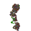
Journal: Dev Cell / Year: 2014 Title: A PH domain in ACAP1 possesses key features of the BAR domain in promoting membrane curvature. Authors: Xiaoyun Pang / Jun Fan / Yan Zhang / Kai Zhang / Bingquan Gao / Jun Ma / Jian Li / Yuchen Deng / Qiangjun Zhou / Edward H Egelman / Victor W Hsu / Fei Sun / Abstract: The BAR (Bin-Amphiphysin-Rvs) domain undergoes dimerization to produce a curved protein structure, which superimposes onto membrane through electrostatic interactions to sense and impart membrane ...The BAR (Bin-Amphiphysin-Rvs) domain undergoes dimerization to produce a curved protein structure, which superimposes onto membrane through electrostatic interactions to sense and impart membrane curvature. In some cases, a BAR domain also possesses an amphipathic helix that inserts into the membrane to induce curvature. ACAP1 (Arfgap with Coil coil, Ankyrin repeat, and PH domain protein 1) contains a BAR domain. Here, we show that this BAR domain can neither bind membrane nor impart curvature, but instead requires a neighboring PH (Pleckstrin Homology) domain to achieve these functions. Specific residues within the PH domain are responsible for both membrane binding and curvature generation. The BAR domain adjacent to the PH domain instead interacts with the BAR domains of neighboring ACAP1 proteins to enable clustering at the membrane. Thus, we have uncovered the molecular basis for an unexpected and unconventional collaboration between PH and BAR domains in membrane bending.
Resolution: 2.2→2.24 Å / Redundancy: 3.2 % / Rmerge(I) obs: 0.418 / Mean I/σ(I) obs: 3.96 / % possible all: 95.6
-
Processing
Software
Name
Version
Classification
ADSC
Quantum
datacollection
SOLVE
phasing
RESOLVE
modelbuilding
OASIS
modelbuilding
REFMAC
5.5.0088
refinement
HKL-2000
datareduction
HKL-2000
datascaling
RESOLVE
phasing
OASIS
phasing
Refinement
Method to determine structure: SAD / Resolution: 2.2→29.85 Å / Cor.coef. Fo:Fc: 0.944 / Cor.coef. Fo:Fc free: 0.914 / SU B: 15.9 / SU ML: 0.179 / Cross valid method: THROUGHOUT / σ(F): 0 / ESU R: 0.302 / ESU R Free: 0.236 / Stereochemistry target values: MAXIMUM LIKELIHOOD / Details: HYDROGENS HAVE BEEN ADDED IN THE RIDING POSITIONS
Rfactor
Num. reflection
% reflection
Selection details
Rfree
0.266
2059
5 %
RANDOM
Rwork
0.211
-
-
-
all
0.214
41114
-
-
obs
0.214
39141
95.2 %
-
Solvent computation
Ion probe radii: 0.8 Å / Shrinkage radii: 0.8 Å / VDW probe radii: 1.2 Å / Solvent model: MASK
Displacement parameters
Biso mean: 56.373 Å2
Baniso -1
Baniso -2
Baniso -3
1-
-1.57 Å2
0 Å2
-2.2 Å2
2-
-
3.62 Å2
0 Å2
3-
-
-
-2.15 Å2
Refinement step
Cycle: LAST / Resolution: 2.2→29.85 Å
Protein
Nucleic acid
Ligand
Solvent
Total
Num. atoms
5839
0
0
146
5985
Refine LS restraints
Refine-ID
Type
Dev ideal
Dev ideal target
Number
X-RAY DIFFRACTION
r_bond_refined_d
0.013
0.022
5935
X-RAY DIFFRACTION
r_angle_refined_deg
1.459
1.968
7986
X-RAY DIFFRACTION
r_dihedral_angle_1_deg
6.026
5
728
X-RAY DIFFRACTION
r_dihedral_angle_2_deg
36.279
23.186
295
X-RAY DIFFRACTION
r_dihedral_angle_3_deg
20.492
15
1114
X-RAY DIFFRACTION
r_dihedral_angle_4_deg
19.053
15
66
X-RAY DIFFRACTION
r_chiral_restr
0.102
0.2
884
X-RAY DIFFRACTION
r_gen_planes_refined
0.006
0.021
4461
X-RAY DIFFRACTION
r_mcbond_it
0.67
1.5
3630
X-RAY DIFFRACTION
r_mcangle_it
1.284
2
5808
X-RAY DIFFRACTION
r_scbond_it
2.227
3
2305
X-RAY DIFFRACTION
r_scangle_it
3.728
4.5
2178
LS refinement shell
Resolution: 2.2→2.26 Å / Total num. of bins used: 20
Rfactor
Num. reflection
% reflection
Rfree
0.339
156
-
Rwork
0.278
2649
-
obs
-
-
89.25 %
Refinement TLS params.
Method: refined / Refine-ID: X-RAY DIFFRACTION
ID
L11 (°2)
L12 (°2)
L13 (°2)
L22 (°2)
L23 (°2)
L33 (°2)
S11 (Å °)
S12 (Å °)
S13 (Å °)
S21 (Å °)
S22 (Å °)
S23 (Å °)
S31 (Å °)
S32 (Å °)
S33 (Å °)
T11 (Å2)
T12 (Å2)
T13 (Å2)
T22 (Å2)
T23 (Å2)
T33 (Å2)
Origin x (Å)
Origin y (Å)
Origin z (Å)
1
7.4414
-0.9379
4.1793
0.5072
-0.3464
3.1095
0.0591
-0.3899
0.0035
-0.002
-0.0532
0.0088
-0.0058
-0.1774
-0.0059
0.1129
-0.0064
0.1
0.0912
0.0158
0.0986
-15.6157
-1.3411
32.4278
2
4.717
-1.4637
2.096
0.7811
-0.6544
1.594
0.0938
-0.3448
-0.0895
0.0648
-0.0007
-0.0349
0.1678
0.0325
-0.0931
0.1822
-0.0125
0.0583
0.1512
0.0374
0.1303
11.0239
-8.3792
46.2037
3
5.8697
0.081
-0.5761
4.9024
-3.4347
10.3702
0.1478
0.0479
-0.1296
-0.1963
-0.1195
-0.0396
0.729
0.4054
-0.0283
0.5358
0.1038
-0.1365
0.2275
0.0343
0.2271
40.2939
-35.9722
82.2715
4
2.2589
1.4655
0.1477
9.0011
2.6397
4.0609
-0.4353
0.444
0.5067
-1.4671
0.0217
0.6167
-0.8487
-0.1075
0.4135
0.5184
0.1013
-0.1676
0.285
0.0224
0.3011
-46.8866
2.0681
-12.8652
5
1.4967
-0.2835
0.968
0.2283
-0.1566
0.6509
0.0936
-0.0085
0.0946
-0.0411
-0.1233
-0.0364
0.1058
-0.0015
0.0297
0.2339
0.0089
0.0963
0.3688
0.0468
0.2676
-9.5988
-5.6709
31.8679
Refinement TLS group
ID
Refine-ID
Refine TLS-ID
Auth asym-ID
Auth seq-ID
1
X-RAY DIFFRACTION
1
A
-3 - 250
2
X-RAY DIFFRACTION
2
B
0 - 250
3
X-RAY DIFFRACTION
3
A
251 - 364
4
X-RAY DIFFRACTION
4
B
251 - 361
5
X-RAY DIFFRACTION
5
A
401 - 479
6
X-RAY DIFFRACTION
5
B
401 - 467
+
About Yorodumi
-
News
-
Feb 9, 2022. New format data for meta-information of EMDB entries
New format data for meta-information of EMDB entries
Version 3 of the EMDB header file is now the official format.
The previous official version 1.9 will be removed from the archive.
In the structure databanks used in Yorodumi, some data are registered as the other names, "COVID-19 virus" and "2019-nCoV". Here are the details of the virus and the list of structure data.
Jan 31, 2019. EMDB accession codes are about to change! (news from PDBe EMDB page)
EMDB accession codes are about to change! (news from PDBe EMDB page)
The allocation of 4 digits for EMDB accession codes will soon come to an end. Whilst these codes will remain in use, new EMDB accession codes will include an additional digit and will expand incrementally as the available range of codes is exhausted. The current 4-digit format prefixed with “EMD-” (i.e. EMD-XXXX) will advance to a 5-digit format (i.e. EMD-XXXXX), and so on. It is currently estimated that the 4-digit codes will be depleted around Spring 2019, at which point the 5-digit format will come into force.
The EM Navigator/Yorodumi systems omit the EMD- prefix.
Related info.:Q: What is EMD? / ID/Accession-code notation in Yorodumi/EM Navigator
Yorodumi is a browser for structure data from EMDB, PDB, SASBDB, etc.
This page is also the successor to EM Navigator detail page, and also detail information page/front-end page for Omokage search.
The word "yorodu" (or yorozu) is an old Japanese word meaning "ten thousand". "mi" (miru) is to see.
Related info.:EMDB / PDB / SASBDB / Comparison of 3 databanks / Yorodumi Search / Aug 31, 2016. New EM Navigator & Yorodumi / Yorodumi Papers / Jmol/JSmol / Function and homology information / Changes in new EM Navigator and Yorodumi
 Movie
Movie Controller


 Open data
 Basic information
 Components
 Keywords
 Function and homology information
 Homo sapiens (human)
 X-RAY DIFFRACTION /
  Authors
 Citation

 Structure visualization
 Downloads & links
 Other downloads


 PDBj
PDBj

 Assembly
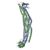

 Mass: 18.015 Da / Num. of mol.: 146 / Source method: isolated from a natural source / Formula: H2O
 Sample preparation
 Processing