 Movie
Movie Controller


+ Open data

- Basic information
| Entry | Database: PDB / ID: 6xgq | ||||||||||||
|---|---|---|---|---|---|---|---|---|---|---|---|---|---|
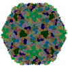
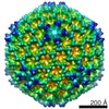
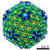
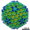
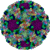
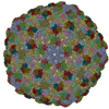
| Title | YSD1 bacteriophage capsid | ||||||||||||
 Components Components |
| ||||||||||||
 Keywords Keywords | VIRUS / Bacteriophage capsid | ||||||||||||
| Function / homology |  Function and homology information Function and homology information | ||||||||||||
| Biological species |  Bacteriophage sp. (virus) Bacteriophage sp. (virus) | ||||||||||||
| Method | ELECTRON MICROSCOPY / single particle reconstruction / cryo EM / Resolution: 3.8 Å | ||||||||||||
 Authors Authors | Hardy, J.M. / Dunstan, R. / Venugopal, H. / Lithgow, T.J. / Coulibaly, F.J. | ||||||||||||
| Funding support |  Australia, Australia,  United Kingdom, 3items United Kingdom, 3items
| ||||||||||||
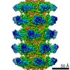
 Citation Citation | Journal: Nat Commun / Year: 2020 Title: The architecture and stabilisation of flagellotropic tailed bacteriophages. Authors: Joshua M Hardy / Rhys A Dunstan / Rhys Grinter / Matthew J Belousoff / Jiawei Wang / Derek Pickard / Hariprasad Venugopal / Gordon Dougan / Trevor Lithgow / Fasséli Coulibaly / Abstract: Flagellotropic bacteriophages engage flagella to reach the bacterial surface as an effective means to increase the capture radius for predation. Structural details of these viruses are of great ...Flagellotropic bacteriophages engage flagella to reach the bacterial surface as an effective means to increase the capture radius for predation. Structural details of these viruses are of great interest given the substantial drag forces and torques they face when moving down the spinning flagellum. We show that the main capsid and auxiliary proteins form two nested chainmails that ensure the integrity of the bacteriophage head. Core stabilising structures are conserved in herpesviruses suggesting their ancestral origin. The structure of the tail also reveals a robust yet pliable assembly. Hexameric rings of the tail-tube protein are braced by the N-terminus and a β-hairpin loop, and interconnected along the tail by the splayed β-hairpins. By contrast, we show that the β-hairpin has an inhibitory role in the tail-tube precursor, preventing uncontrolled self-assembly. Dyads of acidic residues inside the tail-tube present regularly-spaced motifs well suited to DNA translocation into bacteria through the tail. | ||||||||||||
| History |
|
- Structure visualization
| Movie |
Movie viewer |
|---|---|
| Structure viewer | Molecule: MolmilJmol/JSmol |
- Downloads & links
-Download
| PDBx/mmCIF format | 6xgq.cif.gz | 551.1 KB | Display | PDBx/mmCIF format |
|---|---|---|---|---|
| PDB format | pdb6xgq.ent.gz | 463.3 KB | Display | PDB format |
| PDBx/mmJSON format | 6xgq.json.gz | Tree view | PDBx/mmJSON format | |
| Others |  Other downloads Other downloads |
-Validation report
| Summary document | 6xgq_validation.pdf.gz | 1.5 MB | Display | wwPDB validaton report |
|---|---|---|---|---|
| Full document | 6xgq_full_validation.pdf.gz | 1.5 MB | Display | |
| Data in XML | 6xgq_validation.xml.gz | 99.8 KB | Display | |
| Data in CIF | 6xgq_validation.cif.gz | 155.9 KB | Display | |
| Arichive directory | https://data.pdbj.org/pub/pdb/validation_reports/xg/6xgqftp://data.pdbj.org/pub/pdb/validation_reports/xg/6xgq | HTTPS FTP |
-Related structure data
| Related structure data | 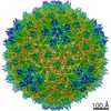 22182MC  6xgpC  6xgrC M: map data used to model this data C: citing same article ( |
|---|---|
| Similar structure data |

-Links
 PDBj
PDBj- Assembly
| Deposited unit | 
|
|---|---|
| 1 | x 60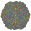
|
| 2 |
|
| 3 | x 5
|
| 4 | x 6
|
| 5 | 
|
| Symmetry | Point symmetry: (Schoenflies symbol: I (icosahedral)) |
-Components
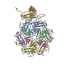
| #1: Protein | Mass: 39945.598 Da / Num. of mol.: 7 / Source method: isolated from a natural source / Source: (natural) Bacteriophage sp. (virus) / References: UniProt: A0A498U580#2: Protein | Mass: 14401.047 Da / Num. of mol.: 7 / Source method: isolated from a natural source / Source: (natural) Bacteriophage sp. (virus) / References: UniProt: A0A498TZZ8 |
|---|
-Experimental details
-Experiment
| Experiment | Method: ELECTRON MICROSCOPY |
|---|---|
| EM experiment | Aggregation state: PARTICLE / 3D reconstruction method: single particle reconstruction |
- Sample preparation
| Component | Name: Bacteriophage sp. / Type: VIRUS Details: From environmental water samples taken during a phage survey of the waterways of Cambridge UK, the phage YSD1 was isolated using the attenuated S. enterica serovar Typhi BRD948. The virus ...Details: From environmental water samples taken during a phage survey of the waterways of Cambridge UK, the phage YSD1 was isolated using the attenuated S. enterica serovar Typhi BRD948. The virus was then amplified by infecting S. Typhimurium SL3261 delta-fljB. Entity ID: all / Source: NATURAL | ||||||||||||||||||||
|---|---|---|---|---|---|---|---|---|---|---|---|---|---|---|---|---|---|---|---|---|---|
| Molecular weight | Value: 22.8 MDa | ||||||||||||||||||||
| Source (natural) | Organism: Bacteriophage sp. (virus) / Strain: YSD1 | ||||||||||||||||||||
| Details of virus | Empty: NO / Enveloped: NO / Isolate: SUBSPECIES / Type: VIRION | ||||||||||||||||||||
| Natural host | Organism: Salmonella enterica subsp. enterica serovar Typhi | ||||||||||||||||||||
| Virus shell | Name: YSD1 capsid / Diameter: 650 nm / Triangulation number (T number): 7 | ||||||||||||||||||||
| Buffer solution | pH: 7.5 | ||||||||||||||||||||
| Buffer component |
| ||||||||||||||||||||
| Specimen | Embedding applied: NO / Shadowing applied: NO / Staining applied: NO / Vitrification applied: YES | ||||||||||||||||||||
| Vitrification | Instrument: FEI VITROBOT MARK IV / Cryogen name: ETHANE / Humidity: 100 % / Chamber temperature: 277 K Details: The grid was blotted for 2 seconds with a blot force of -3 and no drain time. |
- Electron microscopy imaging
| Experimental equipment |  Model: Titan Krios / Image courtesy: FEI Company |
|---|---|
| Microscopy | Model: FEI TITAN KRIOS |
| Electron gun | Electron source:  FIELD EMISSION GUN / Accelerating voltage: 300 kV / Illumination mode: FLOOD BEAM FIELD EMISSION GUN / Accelerating voltage: 300 kV / Illumination mode: FLOOD BEAM |
| Electron lens | Mode: BRIGHT FIELD / Calibrated magnification: 105000 X / Calibrated defocus min: 500 nm / Calibrated defocus max: 2500 nm / Cs: 2.7 mm |
| Specimen holder | Cryogen: NITROGEN / Specimen holder model: FEI TITAN KRIOS AUTOGRID HOLDER |
| Image recording | Average exposure time: 12 sec. / Electron dose: 27.24 e/Å2 / Detector mode: SUPER-RESOLUTION / Film or detector model: GATAN K2 QUANTUM (4k x 4k) / Num. of grids imaged: 1 / Num. of real images: 1881 |
| Image scans | Movie frames/image: 30 / Used frames/image: 1-30 |
- Processing
| EM software |
| |||||||||||||||||||||||||||||||||||||||||||||
|---|---|---|---|---|---|---|---|---|---|---|---|---|---|---|---|---|---|---|---|---|---|---|---|---|---|---|---|---|---|---|---|---|---|---|---|---|---|---|---|---|---|---|---|---|---|---|
| CTF correction | Type: PHASE FLIPPING AND AMPLITUDE CORRECTION | |||||||||||||||||||||||||||||||||||||||||||||
| Particle selection | Num. of particles selected: 7366 | |||||||||||||||||||||||||||||||||||||||||||||
| Symmetry | Point symmetry: I (icosahedral) | |||||||||||||||||||||||||||||||||||||||||||||
| 3D reconstruction | Resolution: 3.8 Å / Resolution method: FSC 0.143 CUT-OFF / Num. of particles: 5449 / Algorithm: BACK PROJECTION / Num. of class averages: 11 / Symmetry type: POINT | |||||||||||||||||||||||||||||||||||||||||||||
| Atomic model building | Protocol: RIGID BODY FIT / Space: REAL | |||||||||||||||||||||||||||||||||||||||||||||
| Atomic model building | PDB-ID: 6XGP Pdb chain-ID: A / Accession code: 6XGP / Source name: PDB / Type: experimental model |
