 Movie
Movie Controller

[English] 日本語
 Yorodumi
- PDB-6rf2: Cryo-EM structure of the C-terminal DC repeat (CDC) of human doub... -
+ Open data

- Basic information
| Entry | Database: PDB / ID: 6rf2 | ||||||
|---|---|---|---|---|---|---|---|
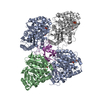
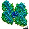
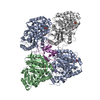
| Title | Cryo-EM structure of the C-terminal DC repeat (CDC) of human doublecortin (DCX) bound to 13-protofilament GDP.Pi-microtubule | ||||||
 Components Components |
| ||||||
 Keywords Keywords | CYTOSOLIC PROTEIN / Microtubule-associated protein / neuronal migration protein / microtubule nucleation and stabilisation | ||||||
| Function / homology |  Function and homology information Function and homology informationaxoneme assembly / Neurofascin interactions / positive regulation of axon guidance / microtubule associated complex / microtubule-based process / cytoplasmic microtubule / cellular response to interleukin-4 / central nervous system development / structural constituent of cytoskeleton / microtubule cytoskeleton organization ...axoneme assembly / Neurofascin interactions / positive regulation of axon guidance / microtubule associated complex / microtubule-based process / cytoplasmic microtubule / cellular response to interleukin-4 / central nervous system development / structural constituent of cytoskeleton / microtubule cytoskeleton organization / neuron migration / nervous system development / mitotic cell cycle / double-stranded RNA binding / microtubule cytoskeleton / retina development in camera-type eye / microtubule binding / cytoskeleton / microtubule / Hydrolases; Acting on acid anhydrides; Acting on GTP to facilitate cellular and subcellular movement / neuron projection / intracellular signal transduction / cilium / protein heterodimerization activity / GTPase activity / ubiquitin protein ligase binding / protein kinase binding / GTP binding / metal ion binding / cytoplasm / cytosol Similarity search - Function | ||||||
| Biological species |  Homo sapiens (human) Homo sapiens (human) | ||||||
| Method | ELECTRON MICROSCOPY / single particle reconstruction / cryo EM / Resolution: 4.2 Å | ||||||
 Authors Authors | Manka, S.W. | ||||||
| Funding support |  United Kingdom, 1items United Kingdom, 1items
| ||||||
 Citation Citation | Journal: EMBO Rep / Year: 2020 Title: Pseudo-repeats in doublecortin make distinct mechanistic contributions to microtubule regulation. Authors: Szymon W Manka / Carolyn A Moores / Abstract: Doublecortin (DCX) is a neuronal microtubule-associated protein (MAP) indispensable for brain development. Its flexibly linked doublecortin (DC) domains-NDC and CDC-mediate microtubule (MT) ...Doublecortin (DCX) is a neuronal microtubule-associated protein (MAP) indispensable for brain development. Its flexibly linked doublecortin (DC) domains-NDC and CDC-mediate microtubule (MT) nucleation and stabilization, but it is unclear how. Using high-resolution time-resolved cryo-EM, we mapped NDC and CDC interactions with tubulin at different MT polymerization stages and studied their functional effects on MT dynamics using TIRF microscopy. Although coupled, each DC repeat within DCX appears to have a distinct role in MT nucleation and stabilization: CDC is a conformationally plastic module that appears to facilitate MT nucleation and stabilize tubulin-tubulin contacts in the nascent MT lattice, while NDC appears to be favored along the mature lattice, providing MT stabilization. Our structures of MT-bound DC domains also explain in unprecedented detail the DCX mutation-related brain defects observed in the clinic. This modular composition of DCX reflects a common design principle among MAPs where pseudo-repeats of tubulin/MT binding elements chaperone or stabilize distinct conformational transitions to regulate distinct stages of MT dynamic instability. | ||||||
| History |
|
- Structure visualization
| Movie |
Movie viewer |
|---|---|
| Structure viewer | Molecule: MolmilJmol/JSmol |
- Downloads & links
-Download
| PDBx/mmCIF format | 6rf2.cif.gz | 323.2 KB | Display | PDBx/mmCIF format |
|---|---|---|---|---|
| PDB format | pdb6rf2.ent.gz | 259.4 KB | Display | PDB format |
| PDBx/mmJSON format | 6rf2.json.gz | Tree view | PDBx/mmJSON format | |
| Others |  Other downloads Other downloads |
-Validation report
| Arichive directory | https://data.pdbj.org/pub/pdb/validation_reports/rf/6rf2ftp://data.pdbj.org/pub/pdb/validation_reports/rf/6rf2 | HTTPS FTP |
|---|
-Related structure data
| Related structure data |  4861MC  4858C 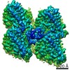 4863C  6revC  6rfdC M: map data used to model this data C: citing same article ( |
|---|---|
| Similar structure data |


-Links
 PDBj
PDBj


- Assembly
| Deposited unit | 
|
|---|---|
| 1 |
|
-Components
-Protein , 3 types, 5 molecules aABbC
| #1: Protein | Mass: 48263.594 Da / Num. of mol.: 2 / Source method: isolated from a natural source / Source: (natural) #2: Protein | Mass: 48113.129 Da / Num. of mol.: 2 / Source method: isolated from a natural source / Source: (natural) #3: Protein | | Mass: 9900.500 Da / Num. of mol.: 1 Source method: isolated from a genetically manipulated source Source: (gene. exp.) Homo sapiens (human) / Gene: DCX, DBCN, LISX / Production host:  |
|---|
-Non-polymers , 4 types, 8 molecules 


| #4: Chemical |  Mass: 523.180 Da / Num. of mol.: 2 / Source method: obtained synthetically / Formula: C10H16N5O14P3 / Comment: GTP, energy-carrying molecule*YM Mass: 523.180 Da / Num. of mol.: 2 / Source method: obtained synthetically / Formula: C10H16N5O14P3 / Comment: GTP, energy-carrying molecule*YM#5: Chemical |  Mass: 24.305 Da / Num. of mol.: 2 / Source method: obtained synthetically / Formula: Mg Mass: 24.305 Da / Num. of mol.: 2 / Source method: obtained synthetically / Formula: Mg#6: Chemical |  Mass: 94.971 Da / Num. of mol.: 2 / Source method: obtained synthetically / Formula: PO4 Mass: 94.971 Da / Num. of mol.: 2 / Source method: obtained synthetically / Formula: PO4#7: Chemical |  Type: RNA linking / Mass: 443.201 Da / Num. of mol.: 2 / Source method: obtained synthetically / Formula: C10H15N5O11P2 / Comment: GDP, energy-carrying molecule*YM Type: RNA linking / Mass: 443.201 Da / Num. of mol.: 2 / Source method: obtained synthetically / Formula: C10H15N5O11P2 / Comment: GDP, energy-carrying molecule*YM |
|---|
-Experimental details
-Experiment
| Experiment | Method: ELECTRON MICROSCOPY |
|---|---|
| EM experiment | Aggregation state: FILAMENT / 3D reconstruction method: single particle reconstruction |
- Sample preparation
| Component |
| ||||||||||||||||||||||||
|---|---|---|---|---|---|---|---|---|---|---|---|---|---|---|---|---|---|---|---|---|---|---|---|---|---|
| Source (natural) |
| ||||||||||||||||||||||||
| Source (recombinant) | Organism: | ||||||||||||||||||||||||
| Buffer solution | pH: 6.8 | ||||||||||||||||||||||||
| Specimen | Embedding applied: NO / Shadowing applied: NO / Staining applied: NO / Vitrification applied: YES | ||||||||||||||||||||||||
| Vitrification | Cryogen name: ETHANE |
- Electron microscopy imaging
| Experimental equipment |  Model: Tecnai Polara / Image courtesy: FEI Company |
|---|---|
| Microscopy | Model: FEI POLARA 300 |
| Electron gun | Electron source:  FIELD EMISSION GUN / Accelerating voltage: 300 kV / Illumination mode: FLOOD BEAM FIELD EMISSION GUN / Accelerating voltage: 300 kV / Illumination mode: FLOOD BEAM |
| Electron lens | Mode: BRIGHT FIELD |
| Image recording | Electron dose: 25 e/Å2 / Film or detector model: GATAN K2 SUMMIT (4k x 4k) |
- Processing
| CTF correction | Type: PHASE FLIPPING AND AMPLITUDE CORRECTION |
|---|---|
| 3D reconstruction | Resolution: 4.2 Å / Resolution method: FSC 0.143 CUT-OFF / Num. of particles: 6591 / Symmetry type: POINT |
| Atomic model building | PDB-ID: 5IP4 Accession code: 5IP4 / Source name: PDB / Type: experimental model |

