Movie
Movie Controller

[English] 日本語
 Yorodumi
- PDB-6rf8: Cryo-EM structure of the N-terminal DC repeat (NDC) of NDC-NDC ch... -
+ Open data

- Basic information
| Entry | Database: PDB / ID: 6rf8 | ||||||
|---|---|---|---|---|---|---|---|
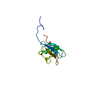
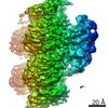
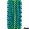
| Title | Cryo-EM structure of the N-terminal DC repeat (NDC) of NDC-NDC chimera (human sequence) bound to 13-protofilament GDP-microtubule | ||||||
 Components Components |
| ||||||
 Keywords Keywords | CYTOSOLIC PROTEIN / Microtubule-associated protein / neuronal migration protein / ubiquitin-like fold / microtubule nucleation and stabilisation | ||||||
| Function / homology |  Function and homology information Function and homology informationaxoneme assembly / Neurofascin interactions / positive regulation of axon guidance / microtubule associated complex / microtubule-based process / cytoplasmic microtubule / cellular response to interleukin-4 / central nervous system development / structural constituent of cytoskeleton / microtubule cytoskeleton organization ...axoneme assembly / Neurofascin interactions / positive regulation of axon guidance / microtubule associated complex / microtubule-based process / cytoplasmic microtubule / cellular response to interleukin-4 / central nervous system development / structural constituent of cytoskeleton / microtubule cytoskeleton organization / neuron migration / nervous system development / mitotic cell cycle / double-stranded RNA binding / microtubule cytoskeleton / retina development in camera-type eye / microtubule binding / cytoskeleton / microtubule / Hydrolases; Acting on acid anhydrides; Acting on GTP to facilitate cellular and subcellular movement / neuron projection / intracellular signal transduction / cilium / protein heterodimerization activity / GTPase activity / ubiquitin protein ligase binding / protein kinase binding / GTP binding / metal ion binding / cytoplasm / cytosol Similarity search - Function | ||||||
| Biological species |  Homo sapiens (human) Homo sapiens (human) | ||||||
| Method | ELECTRON MICROSCOPY / single particle reconstruction / cryo EM / Resolution: 3.8 Å | ||||||
 Authors Authors | Manka, S.W. | ||||||
| Funding support |  United Kingdom, 1items United Kingdom, 1items
| ||||||
 Citation Citation | Journal: J Struct Biol / Year: 2020 Title: A microtubule RELION-based pipeline for cryo-EM image processing. Authors: Alexander D Cook / Szymon W Manka / Su Wang / Carolyn A Moores / Joseph Atherton / Abstract: Microtubules are polar filaments built from αβ-tubulin heterodimers that exhibit a range of architectures in vitro and in vivo. Tubulin heterodimers are arranged helically in the microtubule wall ...Microtubules are polar filaments built from αβ-tubulin heterodimers that exhibit a range of architectures in vitro and in vivo. Tubulin heterodimers are arranged helically in the microtubule wall but many physiologically relevant architectures exhibit a break in helical symmetry known as the seam. Noisy 2D cryo-electron microscopy projection images of pseudo-helical microtubules therefore depict distinct but highly similar views owing to the high structural similarity of α- and β-tubulin. The determination of the αβ-tubulin register and seam location during image processing is essential for alignment accuracy that enables determination of biologically relevant structures. Here we present a pipeline designed for image processing and high-resolution reconstruction of cryo-electron microscopy microtubule datasets, based in the popular and user-friendly RELION image-processing package, Microtubule RELION-based Pipeline (MiRP). The pipeline uses a combination of supervised classification and prior knowledge about geometric lattice constraints in microtubules to accurately determine microtubule architecture and seam location. The presented method is fast and semi-automated, producing near-atomic resolution reconstructions with test datasets that contain a range of microtubule architectures and binding proteins. | ||||||
| History |
|
- Structure visualization
| Movie |
Movie viewer |
|---|---|
| Structure viewer | Molecule: MolmilJmol/JSmol |
- Downloads & links
-Download
| PDBx/mmCIF format | 6rf8.cif.gz | 326.3 KB | Display | PDBx/mmCIF format |
|---|---|---|---|---|
| PDB format | pdb6rf8.ent.gz | 261.7 KB | Display | PDB format |
| PDBx/mmJSON format | 6rf8.json.gz | Tree view | PDBx/mmJSON format | |
| Others |  Other downloads Other downloads |
-Validation report
| Arichive directory | https://data.pdbj.org/pub/pdb/validation_reports/rf/6rf8ftp://data.pdbj.org/pub/pdb/validation_reports/rf/6rf8 | HTTPS FTP |
|---|
-Related structure data
| Related structure data | 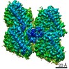 4862MC M: map data used to model this data C: citing same article ( |
|---|---|
| Similar structure data |

-Links
 PDBj
PDBj


- Assembly
| Deposited unit | 
|
|---|---|
| 1 |
|
-Components
-Protein , 3 types, 5 molecules NbBAa
| #1: Protein | Mass: 11441.834 Da / Num. of mol.: 1 Source method: isolated from a genetically manipulated source Source: (gene. exp.) Homo sapiens (human) / Gene: DCX, DBCN, LISX / Production host:  | ||
|---|---|---|---|
| #2: Protein | Mass: 48113.129 Da / Num. of mol.: 2 / Source method: isolated from a natural source / Source: (natural) #3: Protein | Mass: 48263.594 Da / Num. of mol.: 2 / Source method: isolated from a natural source / Source: (natural) |
-Non-polymers , 3 types, 6 molecules 


| #4: Chemical |  Type: RNA linking / Mass: 443.201 Da / Num. of mol.: 2 / Source method: obtained synthetically / Formula: C10H15N5O11P2 / Comment: GDP, energy-carrying molecule*YM Type: RNA linking / Mass: 443.201 Da / Num. of mol.: 2 / Source method: obtained synthetically / Formula: C10H15N5O11P2 / Comment: GDP, energy-carrying molecule*YM#5: Chemical |  Mass: 523.180 Da / Num. of mol.: 2 / Source method: obtained synthetically / Formula: C10H16N5O14P3 / Comment: GTP, energy-carrying molecule*YM Mass: 523.180 Da / Num. of mol.: 2 / Source method: obtained synthetically / Formula: C10H16N5O14P3 / Comment: GTP, energy-carrying molecule*YM#6: Chemical |  Mass: 24.305 Da / Num. of mol.: 2 / Source method: obtained synthetically / Formula: Mg Mass: 24.305 Da / Num. of mol.: 2 / Source method: obtained synthetically / Formula: Mg |
|---|
-Experimental details
-Experiment
| Experiment | Method: ELECTRON MICROSCOPY |
|---|---|
| EM experiment | Aggregation state: FILAMENT / 3D reconstruction method: single particle reconstruction |
- Sample preparation
| Component |
| ||||||||||||||||||||||||
|---|---|---|---|---|---|---|---|---|---|---|---|---|---|---|---|---|---|---|---|---|---|---|---|---|---|
| Source (natural) |
| ||||||||||||||||||||||||
| Source (recombinant) | Organism: | ||||||||||||||||||||||||
| Buffer solution | pH: 6.8 | ||||||||||||||||||||||||
| Specimen | Embedding applied: NO / Shadowing applied: NO / Staining applied: NO / Vitrification applied: YES | ||||||||||||||||||||||||
| Vitrification | Cryogen name: ETHANE |
- Electron microscopy imaging
| Experimental equipment |  Model: Tecnai Polara / Image courtesy: FEI Company |
|---|---|
| Microscopy | Model: FEI POLARA 300 |
| Electron gun | Electron source:  FIELD EMISSION GUN / Accelerating voltage: 300 kV / Illumination mode: FLOOD BEAM FIELD EMISSION GUN / Accelerating voltage: 300 kV / Illumination mode: FLOOD BEAM |
| Electron lens | Mode: BRIGHT FIELD |
| Image recording | Electron dose: 25 e/Å2 / Detector mode: COUNTING / Film or detector model: GATAN K2 SUMMIT (4k x 4k) |
- Processing
| EM software | Name: PHENIX / Category: model fitting |
|---|---|
| CTF correction | Type: PHASE FLIPPING AND AMPLITUDE CORRECTION |
| 3D reconstruction | Resolution: 3.8 Å / Resolution method: FSC 0.143 CUT-OFF / Num. of particles: 28347 / Symmetry type: POINT |
| Atomic model building | Protocol: FLEXIBLE FIT / Space: REAL / Target criteria: Cross-correlation coefficient |
| Atomic model building | PDB-ID: 1MJD Accession code: 1MJD / Source name: PDB / Type: experimental model |