 Movie
Movie Controller

[English] 日本語
 Yorodumi
- EMDB-10195: Doublecortin N-terminal DC-domain (NDC)-decorated 13-protofilamen... -
+ Open data

- Basic information
| Entry | Database: EMDB / ID: EMD-10195 | ||||||||||||||||||
|---|---|---|---|---|---|---|---|---|---|---|---|---|---|---|---|---|---|---|---|
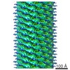
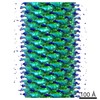
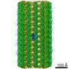
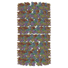
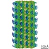
| Title | Doublecortin N-terminal DC-domain (NDC)-decorated 13-protofilament microtubule (GDP state) processed with MiRP (Microtubule Relion-based Pipeline) | ||||||||||||||||||
 Map data Map data | C1 reconstruction of 13-protofilament GDP-microtubule decorated with NDC-NDC chimera of human doublecortin | ||||||||||||||||||
 Sample Sample |
| ||||||||||||||||||
| Biological species |  Homo sapiens (human) Homo sapiens (human) | ||||||||||||||||||
| Method | helical reconstruction / cryo EM / Resolution: 4.5 Å | ||||||||||||||||||
 Authors Authors | Cook AC / Manka SW / Wang S / Moores CA / Atherton J | ||||||||||||||||||
| Funding support |  United Kingdom, 5 items United Kingdom, 5 items
| ||||||||||||||||||
 Citation Citation | Journal: J Struct Biol / Year: 2020 Title: A microtubule RELION-based pipeline for cryo-EM image processing. Authors: Alexander D Cook / Szymon W Manka / Su Wang / Carolyn A Moores / Joseph Atherton / Abstract: Microtubules are polar filaments built from αβ-tubulin heterodimers that exhibit a range of architectures in vitro and in vivo. Tubulin heterodimers are arranged helically in the microtubule wall ...Microtubules are polar filaments built from αβ-tubulin heterodimers that exhibit a range of architectures in vitro and in vivo. Tubulin heterodimers are arranged helically in the microtubule wall but many physiologically relevant architectures exhibit a break in helical symmetry known as the seam. Noisy 2D cryo-electron microscopy projection images of pseudo-helical microtubules therefore depict distinct but highly similar views owing to the high structural similarity of α- and β-tubulin. The determination of the αβ-tubulin register and seam location during image processing is essential for alignment accuracy that enables determination of biologically relevant structures. Here we present a pipeline designed for image processing and high-resolution reconstruction of cryo-electron microscopy microtubule datasets, based in the popular and user-friendly RELION image-processing package, Microtubule RELION-based Pipeline (MiRP). The pipeline uses a combination of supervised classification and prior knowledge about geometric lattice constraints in microtubules to accurately determine microtubule architecture and seam location. The presented method is fast and semi-automated, producing near-atomic resolution reconstructions with test datasets that contain a range of microtubule architectures and binding proteins. | ||||||||||||||||||
| History |
|
- Structure visualization
| Movie |
 Movie viewer Movie viewer |
|---|---|
| Structure viewer | EM map: SurfViewMolmilJmol/JSmol |
| Supplemental images |

- Downloads & links
-EMDB archive
| Map data | emd_10195.map.gz | 245.8 MB | EMDB map data format | |
|---|---|---|---|---|
| Header (meta data) | emd-10195-v30.xmlemd-10195.xml | 11.7 KB 11.7 KB | Display Display | EMDB header |
| Images | 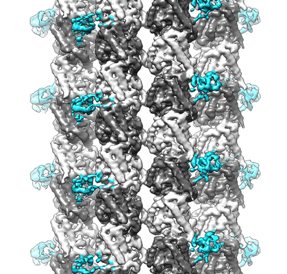 emd_10195.png emd_10195.png | 229.9 KB | ||
| Others | emd_10195_additional.map.gzemd_10195_additional_1.map.gz | 488 KB 488 KB | ||
| Archive directory |  http://ftp.pdbj.org/pub/emdb/structures/EMD-10195ftp://ftp.pdbj.org/pub/emdb/structures/EMD-10195 http://ftp.pdbj.org/pub/emdb/structures/EMD-10195ftp://ftp.pdbj.org/pub/emdb/structures/EMD-10195 | HTTPS FTP |
-Related structure data
| Related structure data |  4862C  6rf8C C: citing same article ( |
|---|---|
| Similar structure data | |
| EM raw data | EMPIAR-10300 (Title: Cryo micrographs of microtubules (GDP state) decorated with NDC-NDC chimera of human doublecortin Data size: 50.4 Data #1: Aligned movie frames of microtubules decorated with NDC-NDC chimera of human doublecortin [micrographs - multiframe]) |

-Links
| EMDB pages | EMDB (EBI/PDBe) / EMDataResource |
|---|
-Map
| File | Download / File: emd_10195.map.gz / Format: CCP4 / Size: 307.5 MB / Type: IMAGE STORED AS FLOATING POINT NUMBER (4 BYTES) | ||||||||||||||||||||||||||||||||||||||||||||||||||||||||||||
|---|---|---|---|---|---|---|---|---|---|---|---|---|---|---|---|---|---|---|---|---|---|---|---|---|---|---|---|---|---|---|---|---|---|---|---|---|---|---|---|---|---|---|---|---|---|---|---|---|---|---|---|---|---|---|---|---|---|---|---|---|---|
| Annotation | C1 reconstruction of 13-protofilament GDP-microtubule decorated with NDC-NDC chimera of human doublecortin | ||||||||||||||||||||||||||||||||||||||||||||||||||||||||||||
| Projections & slices | Image control
Images are generated by Spider. | ||||||||||||||||||||||||||||||||||||||||||||||||||||||||||||
| Voxel size | X=Y=Z: 1.39 Å | ||||||||||||||||||||||||||||||||||||||||||||||||||||||||||||
| Density |
| ||||||||||||||||||||||||||||||||||||||||||||||||||||||||||||
| Symmetry | Space group: 1 | ||||||||||||||||||||||||||||||||||||||||||||||||||||||||||||
| Details | EMDB XML:
CCP4 map header:
| ||||||||||||||||||||||||||||||||||||||||||||||||||||||||||||
 Z (Sec.)
 Y (Row.)
 X (Col.)


-Supplemental data
-Additional map: None
| File | emd_10195_additional.map | ||||||||||||
|---|---|---|---|---|---|---|---|---|---|---|---|---|---|
| Annotation | None | ||||||||||||
| Projections & Slices |
| ||||||||||||
| Density Histograms |


-Additional map: None
| File | emd_10195_additional_1.map | ||||||||||||
|---|---|---|---|---|---|---|---|---|---|---|---|---|---|
| Annotation | None | ||||||||||||
| Projections & Slices |
| ||||||||||||
| Density Histograms |


- Sample components
-Entire : 13-protofilament microtubule decorated with NDC-NDC chimera of hu...
| Entire | Name: 13-protofilament microtubule decorated with NDC-NDC chimera of human doublecortin |
|---|---|
| Components |
|
-Supramolecule #1: 13-protofilament microtubule decorated with NDC-NDC chimera of hu...
| Supramolecule | Name: 13-protofilament microtubule decorated with NDC-NDC chimera of human doublecortin type: complex / ID: 1 / Parent: 0 |
|---|---|
| Source (natural) | Organism: Homo sapiens (human) |
| Recombinant expression | Organism:  |
-Experimental details
-Structure determination
| Method | cryo EM |
|---|---|
 Processing Processing | helical reconstruction |
| Aggregation state | filament |
-Sample preparation
| Buffer | pH: 6.8 |
|---|---|
| Vitrification | Cryogen name: ETHANE |
- Electron microscopy
| Microscope | FEI POLARA 300 |
|---|---|
| Image recording | Film or detector model: GATAN K2 SUMMIT (4k x 4k) / Average electron dose: 25.0 e/Å2 |
| Electron beam | Acceleration voltage: 300 kV / Electron source:  FIELD EMISSION GUN FIELD EMISSION GUN |
| Electron optics | Illumination mode: FLOOD BEAM / Imaging mode: BRIGHT FIELD |
| Experimental equipment |  Model: Tecnai Polara / Image courtesy: FEI Company |
-Image processing
| Details | Microtubule RELION-based pipeline (MiRP) |
|---|---|
| Final reconstruction | Applied symmetry - Helical parameters - Δz: 9.46 Å Applied symmetry - Helical parameters - Δ&Phi: -27.67 ° Applied symmetry - Helical parameters - Axial symmetry: C1 (asymmetric) Resolution.type: BY AUTHOR / Resolution: 4.5 Å / Resolution method: FSC 0.143 CUT-OFF / Number images used: 33392 |
| Final angle assignment | Type: NOT APPLICABLE |
