 Movie
Movie Controller

[English] 日本語
 Yorodumi
- PDB-6h9c: Cryo-EM structure of archaeal extremophilic internal membrane-con... -
+ Open data

- Basic information
| Entry | Database: PDB / ID: 6h9c | |||||||||||||||
|---|---|---|---|---|---|---|---|---|---|---|---|---|---|---|---|---|
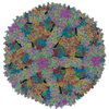
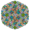
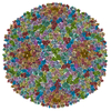
| Title | Cryo-EM structure of archaeal extremophilic internal membrane-containing Haloarcula californiae icosahedral virus 1 (HCIV-1) at 3.74 Angstroms resolution. | |||||||||||||||
 Components Components |
| |||||||||||||||
 Keywords Keywords | VIRUS / vertical single beta-barrel virus / internal membrane-containing archaeal virus. | |||||||||||||||
| Function / homology | VP7 / VP4 / VP9 Function and homology information Function and homology information | |||||||||||||||
| Biological species |  Haloarcula californiae ATCC 33799 (Halophile) Haloarcula californiae ATCC 33799 (Halophile) | |||||||||||||||
| Method | ELECTRON MICROSCOPY / single particle reconstruction / cryo EM / Resolution: 3.74 Å | |||||||||||||||
 Authors Authors | Abrescia, N.G. / Santos-Perez, I. / Charro, D. | |||||||||||||||
| Funding support |  Spain, Spain,  Finland, 4items Finland, 4items
| |||||||||||||||
 Citation Citation | Journal: Nat Commun / Year: 2019 Title: Structural basis for assembly of vertical single β-barrel viruses. Authors: Isaac Santos-Pérez / Diego Charro / David Gil-Carton / Mikel Azkargorta / Felix Elortza / Dennis H Bamford / Hanna M Oksanen / Nicola G A Abrescia / Abstract: The vertical double β-barrel major capsid protein (MCP) fold, fingerprint of the PRD1-adeno viral lineage, is widespread in many viruses infecting organisms across the three domains of life. The ...The vertical double β-barrel major capsid protein (MCP) fold, fingerprint of the PRD1-adeno viral lineage, is widespread in many viruses infecting organisms across the three domains of life. The discovery of PRD1-like viruses with two MCPs challenged the known assembly principles. Here, we present the cryo-electron microscopy (cryo-EM) structures of the archaeal, halophilic, internal membrane-containing Haloarcula californiae icosahedral virus 1 (HCIV-1) and Haloarcula hispanica icosahedral virus 2 (HHIV-2) at 3.7 and 3.8 Å resolution, respectively. Our structures reveal proteins located beneath the morphologically distinct two- and three-tower capsomers and homopentameric membrane proteins at the vertices that orchestrate the positioning of pre-formed vertical single β-barrel MCP heterodimers. The cryo-EM based structures together with the proteomics data provide insights into the assembly mechanism of this type of viruses and into those with membrane-less double β-barrel MCPs. | |||||||||||||||
| History |
|
- Structure visualization
| Movie |
Movie viewer |
|---|---|
| Structure viewer | Molecule: MolmilJmol/JSmol |
- Downloads & links
-Download
| PDBx/mmCIF format | 6h9c.cif.gz | 1.7 MB | Display | PDBx/mmCIF format |
|---|---|---|---|---|
| PDB format | pdb6h9c.ent.gz | 1.4 MB | Display | PDB format |
| PDBx/mmJSON format | 6h9c.json.gz | Tree view | PDBx/mmJSON format | |
| Others |  Other downloads Other downloads |
-Validation report
| Arichive directory | https://data.pdbj.org/pub/pdb/validation_reports/h9/6h9cftp://data.pdbj.org/pub/pdb/validation_reports/h9/6h9c | HTTPS FTP |
|---|
-Related structure data
| Related structure data | 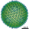 0174MC  0050C  0072C  0073C 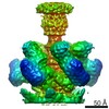 0131C  0172C  6h82C C: citing same article ( M: map data used to model this data |
|---|---|
| Similar structure data |


-Links
 PDBj
PDBj- Assembly
| Deposited unit | 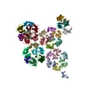
|
|---|---|
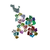
| 1 | x 60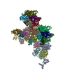
|
-Components
-Protein , 5 types, 30 molecules DFERTSWKJMNILaYbcdZABCPQOVUHGX
| #1: Protein | Mass: 19912.832 Da / Num. of mol.: 15 / Source method: isolated from a natural source Source: (natural) Haloarcula californiae ATCC 33799 (Halophile)Plasmid details: Haloarcula californiae icosahedral virus 1 / References: UniProt: A0A1C7A3R1 #2: Protein | | Mass: 16271.062 Da / Num. of mol.: 1 / Source method: isolated from a natural source Source: (natural) Haloarcula californiae ATCC 33799 (Halophile)References: UniProt: A0A1C7A3R7 #3: Protein | | Mass: 9209.344 Da / Num. of mol.: 1 / Source method: isolated from a natural source Source: (natural) Haloarcula californiae ATCC 33799 (Halophile)#4: Protein | | Mass: 6400.881 Da / Num. of mol.: 1 / Source method: isolated from a natural source Source: (natural) Haloarcula californiae ATCC 33799 (Halophile)Plasmid details: Haloarcula californiae icosahedral virus 1 #7: Protein | Mass: 25995.318 Da / Num. of mol.: 12 / Source method: isolated from a natural source Source: (natural) Haloarcula californiae ATCC 33799 (Halophile)Plasmid details: Haloarcula californiae icosahedral virus 1 / References: UniProt: A0A1C7A3R2 |
|---|
-Protein/peptide , 2 types, 2 molecules ef
| #5: Protein/peptide | ( Mass: 3932.839 Da / Num. of mol.: 1 / Source method: isolated from a natural source Source: (natural) Haloarcula californiae ATCC 33799 (Halophile)Plasmid details: Haloarcula californiae icosahedral virus 1 |
|---|---|
| #6: Protein/peptide | Mass: 1549.902 Da / Num. of mol.: 1 / Source method: isolated from a natural source Source: (natural) Haloarcula californiae ATCC 33799 (Halophile) |
-Experimental details
-Experiment
| Experiment | Method: ELECTRON MICROSCOPY |
|---|---|
| EM experiment | Aggregation state: PARTICLE / 3D reconstruction method: single particle reconstruction |
- Sample preparation
| Component | Name: Haloarcula californiae icosahedral virus 1 / Type: VIRUS / Details: Haloarcula californiae icosahedral virus - 1 / Entity ID: all / Source: NATURAL | ||||||||||||||||||||||||||||||
|---|---|---|---|---|---|---|---|---|---|---|---|---|---|---|---|---|---|---|---|---|---|---|---|---|---|---|---|---|---|---|---|
| Molecular weight | Experimental value: NO | ||||||||||||||||||||||||||||||
| Source (natural) | Organism:  Haloarcula californiae icosahedral virus 1 Haloarcula californiae icosahedral virus 1 | ||||||||||||||||||||||||||||||
| Details of virus | Empty: NO / Enveloped: NO / Isolate: SPECIES / Type: VIRION | ||||||||||||||||||||||||||||||
| Natural host | Organism: Haloarcula californiae ATCC 33799 | ||||||||||||||||||||||||||||||
| Virus shell | Diameter: 800 nm / Triangulation number (T number): 28 | ||||||||||||||||||||||||||||||
| Buffer solution | pH: 7.2 | ||||||||||||||||||||||||||||||
| Buffer component |
| ||||||||||||||||||||||||||||||
| Specimen | Conc.: 1.2 mg/ml / Embedding applied: NO / Shadowing applied: NO / Staining applied: NO / Vitrification applied: YES Details: Haloarcula californiae icosahedral virus 1. Taxonomic identifier 1735722 NCBI. | ||||||||||||||||||||||||||||||
| Vitrification | Instrument: FEI VITROBOT MARK III / Cryogen name: ETHANE |
- Electron microscopy imaging
| Experimental equipment |  Model: Titan Krios / Image courtesy: FEI Company |
|---|---|
| Microscopy | Model: FEI TITAN KRIOS |
| Electron gun | Electron source:  FIELD EMISSION GUN / Accelerating voltage: 300 kV / Illumination mode: FLOOD BEAM FIELD EMISSION GUN / Accelerating voltage: 300 kV / Illumination mode: FLOOD BEAM |
| Electron lens | Mode: BRIGHT FIELD / Nominal defocus max: 3900 nm / Nominal defocus min: 600 nm / Cs: 2.7 mm |
| Specimen holder | Cryogen: NITROGEN / Specimen holder model: FEI TITAN KRIOS AUTOGRID HOLDER |
| Image recording | Electron dose: 36 e/Å2 / Detector mode: INTEGRATING / Film or detector model: FEI FALCON II (4k x 4k) / Num. of grids imaged: 1 / Num. of real images: 3218 |
| Image scans | Movie frames/image: 27 / Used frames/image: 1-26 |
- Processing
| Software | Name: PHENIX / Version: 1.13_2998: / Classification: refinement | |||||||||||||||||||||||||||||||||||||||||||||||||||||||
|---|---|---|---|---|---|---|---|---|---|---|---|---|---|---|---|---|---|---|---|---|---|---|---|---|---|---|---|---|---|---|---|---|---|---|---|---|---|---|---|---|---|---|---|---|---|---|---|---|---|---|---|---|---|---|---|---|
| EM software |
| |||||||||||||||||||||||||||||||||||||||||||||||||||||||
| CTF correction | Type: PHASE FLIPPING AND AMPLITUDE CORRECTION | |||||||||||||||||||||||||||||||||||||||||||||||||||||||
| Particle selection | Num. of particles selected: 4584 | |||||||||||||||||||||||||||||||||||||||||||||||||||||||
| Symmetry | Point symmetry: I (icosahedral) | |||||||||||||||||||||||||||||||||||||||||||||||||||||||
| 3D reconstruction | Resolution: 3.74 Å / Resolution method: FSC 0.143 CUT-OFF / Num. of particles: 3414 / Num. of class averages: 1 / Symmetry type: POINT | |||||||||||||||||||||||||||||||||||||||||||||||||||||||
| Atomic model building | B value: 89.1 / Protocol: AB INITIO MODEL / Space: REAL / Target criteria: Cross-correlation coefficient Details: The HCIV-1 VP7 and VP4 MCPs were manually built aided by beta-barrel core templates derived from the crystal structures of Thermus bacteriophage P23-77 MCPs (PDB ID 3ZN6). | |||||||||||||||||||||||||||||||||||||||||||||||||||||||
| Refinement | Highest resolution: 3.74 Å |
