+ データを開く
- 基本情報
基本情報
| 登録情報 | データベース: PDB / ID: 1pdl | ||||||
|---|---|---|---|---|---|---|---|
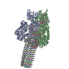
| タイトル | Fitting of gp5 in the cryoEM reconstruction of the bacteriophage T4 baseplate | ||||||
 要素 要素 | Tail-associated lysozyme | ||||||
 キーワード キーワード | HYDROLASE | ||||||
| 機能・相同性 |  機能・相同性情報 機能・相同性情報symbiont entry into host cell via disruption of host cell wall peptidoglycan / virus tail, baseplate / viral tail assembly / symbiont entry into host cell via disruption of host cell envelope / symbiont entry into host / virus tail / peptidoglycan catabolic process / cell wall macromolecule catabolic process / lysozyme / lysozyme activity ...symbiont entry into host cell via disruption of host cell wall peptidoglycan / virus tail, baseplate / viral tail assembly / symbiont entry into host cell via disruption of host cell envelope / symbiont entry into host / virus tail / peptidoglycan catabolic process / cell wall macromolecule catabolic process / lysozyme / lysozyme activity / killing of cells of another organism / defense response to bacterium / symbiont entry into host cell / identical protein binding 類似検索 - 分子機能 | ||||||
| 生物種 |  Enterobacteria phage T4 (ファージ) Enterobacteria phage T4 (ファージ) | ||||||
| 手法 | 電子顕微鏡法 / 単粒子再構成法 / クライオ電子顕微鏡法 / 解像度: 12 Å | ||||||
 データ登録者 データ登録者 | Kostyuchenko, V.A. / Leiman, P.G. / Chipman, P.R. / Kanamaru, S. / van Raaij, M.J. / Arisaka, F. / Mesyanzhinov, V.V. / Rossmann, M.G. | ||||||
 引用 引用 |  ジャーナル: Nat Struct Biol / 年: 2003 ジャーナル: Nat Struct Biol / 年: 2003タイトル: Three-dimensional structure of bacteriophage T4 baseplate. 著者: Victor A Kostyuchenko / Petr G Leiman / Paul R Chipman / Shuji Kanamaru / Mark J van Raaij / Fumio Arisaka / Vadim V Mesyanzhinov / Michael G Rossmann /  要旨: The baseplate of bacteriophage T4 is a multiprotein molecular machine that controls host cell recognition, attachment, tail sheath contraction and viral DNA ejection. We report here the three- ...The baseplate of bacteriophage T4 is a multiprotein molecular machine that controls host cell recognition, attachment, tail sheath contraction and viral DNA ejection. We report here the three-dimensional structure of the baseplate-tail tube complex determined to a resolution of 12 A by cryoelectron microscopy. The baseplate has a six-fold symmetric, dome-like structure approximately 520 A in diameter and approximately 270 A long, assembled around a central hub. A 940 A-long and 96 A-diameter tail tube, coaxial with the hub, is connected to the top of the baseplate. At the center of the dome is a needle-like structure that was previously identified as a cell puncturing device. We have identified the locations of six proteins with known atomic structures, and established the position and shape of several other baseplate proteins. The baseplate structure suggests a mechanism of baseplate triggering and structural transition during the initial stages of T4 infection. #1:  ジャーナル: Nature / 年: 2002 ジャーナル: Nature / 年: 2002タイトル: Structure of the cell-puncturing device of bacteriophage T4 著者: Kanamaru, S. / Leiman, P.G. / Kostyuchenko, V.A. / Chipman, P.R. / Mesyanzhinov, V.V. / Arisaka, F. / Rossmann, M.G. | ||||||
| 履歴 |
| ||||||
| Remark 999 | SEQUENCE COORDINATES FOR CA ATOMS ONLY SUBMITTED. | ||||||
| Remark 300 | BIOMOLECULE: 1 THIS ENTRY CONTAINS A PORTION OF THE BIOLOGICALLY SIGNIFICANT MULTIMER. ASSEMBLY ...BIOMOLECULE: 1 THIS ENTRY CONTAINS A PORTION OF THE BIOLOGICALLY SIGNIFICANT MULTIMER. ASSEMBLY COMPONENTS COM_ID: 1 NAME:GP5 IPR_ID: NULL GO_ID: NULL OTHER_DETAILS: TRIMER |
- 構造の表示
構造の表示
| ムービー |
 ムービービューア ムービービューア |
|---|---|
| 構造ビューア | 分子:  Molmil Molmil Jmol/JSmol Jmol/JSmol |
- ダウンロードとリンク
- ダウンロード
| PDBx/mmCIF形式 |  1pdl.cif.gz 1pdl.cif.gz | 63.1 KB | 表示 |  PDBx/mmCIF形式 PDBx/mmCIF形式 |
|---|---|---|---|---|
| PDB形式 |  pdb1pdl.ent.gz pdb1pdl.ent.gz | 38.9 KB | 表示 |  PDB形式 PDB形式 |
| PDBx/mmJSON形式 |  1pdl.json.gz 1pdl.json.gz | ツリー表示 |  PDBx/mmJSON形式 PDBx/mmJSON形式 | |
| その他 |  その他のダウンロード その他のダウンロード |
-検証レポート
| 文書・要旨 |  1pdl_validation.pdf.gz 1pdl_validation.pdf.gz | 803.4 KB | 表示 |  wwPDB検証レポート wwPDB検証レポート |
|---|---|---|---|---|
| 文書・詳細版 |  1pdl_full_validation.pdf.gz 1pdl_full_validation.pdf.gz | 803 KB | 表示 | |
| XML形式データ |  1pdl_validation.xml.gz 1pdl_validation.xml.gz | 24.8 KB | 表示 | |
| CIF形式データ |  1pdl_validation.cif.gz 1pdl_validation.cif.gz | 36.9 KB | 表示 | |
| アーカイブディレクトリ |  https://data.pdbj.org/pub/pdb/validation_reports/pd/1pdl https://data.pdbj.org/pub/pdb/validation_reports/pd/1pdl ftp://data.pdbj.org/pub/pdb/validation_reports/pd/1pdl ftp://data.pdbj.org/pub/pdb/validation_reports/pd/1pdl | HTTPS FTP |
-関連構造データ
- リンク
リンク
- 集合体
集合体
| 登録構造単位 | 
|
|---|---|
| 1 |
|
| 対称性 | 点対称性: (シェーンフリース記号: C6 (6回回転対称)) |
- 要素
要素
| #1: タンパク質 | 分子量: 63183.723 Da / 分子数: 3 / 由来タイプ: 天然 / 由来: (天然)  Enterobacteria phage T4 (ファージ) / 属: T4-like viruses / 生物種: Enterobacteria phage T4 sensu lato / 参照: UniProt: P16009, lysozyme Enterobacteria phage T4 (ファージ) / 属: T4-like viruses / 生物種: Enterobacteria phage T4 sensu lato / 参照: UniProt: P16009, lysozyme |
|---|
-実験情報
-実験
| 実験 | 手法: 電子顕微鏡法 |
|---|---|
| EM実験 | 試料の集合状態: PARTICLE / 3次元再構成法: 単粒子再構成法 |
- 試料調製
試料調製
| 構成要素 |
| |||||||||||||||
|---|---|---|---|---|---|---|---|---|---|---|---|---|---|---|---|---|
| 緩衝液 | 名称: water / pH: 7 / 詳細: water | |||||||||||||||
| 試料 | 濃度: 5 mg/ml / 包埋: NO / シャドウイング: NO / 染色: NO / 凍結: YES | |||||||||||||||
| 試料支持 | 詳細: holey carbon | |||||||||||||||
| 急速凍結 | 凍結剤: ETHANE / 詳細: ethane vitrification | |||||||||||||||
| 結晶化 | *PLUS 手法: 電子顕微鏡法 |
- 電子顕微鏡撮影
| 顕微鏡 | モデル: FEI/PHILIPS CM300FEG/T / 日付: 2001年1月30日 |
|---|---|
| 電子銃 | 電子線源:  FIELD EMISSION GUN / 加速電圧: 300 kV / 照射モード: FLOOD BEAM FIELD EMISSION GUN / 加速電圧: 300 kV / 照射モード: FLOOD BEAM |
| 電子レンズ | モード: BRIGHT FIELD / 倍率(公称値): 45000 X / 倍率(補正後): 47000 X / 最大 デフォーカス(公称値): 5000 nm / 最小 デフォーカス(公称値): 1200 nm / Cs: 2 mm |
| 試料ホルダ | 温度: 70 K / 傾斜角・最大: 0 ° / 傾斜角・最小: 0 ° |
| 撮影 | 電子線照射量: 25 e/Å2 / フィルム・検出器のモデル: KODAK SO-163 FILM |
- 解析
解析
| EMソフトウェア |
| ||||||||||||
|---|---|---|---|---|---|---|---|---|---|---|---|---|---|
| CTF補正 | 詳細: CTF was corrected for each particle image individually | ||||||||||||
| 対称性 | 点対称性: C6 (6回回転対称) | ||||||||||||
| 3次元再構成 | 手法: model based projection matching / 解像度: 12 Å / 粒子像の数: 945 / ピクセルサイズ(公称値): 3.11 Å / ピクセルサイズ(実測値): 2.98 Å / 倍率補正: TMV images 詳細: a modified version of SPIDER program was used for the reconstruction 対称性のタイプ: POINT | ||||||||||||
| 原子モデル構築 | プロトコル: OTHER / 空間: REAL / Target criteria: Correlation Coefficient maximization / 詳細: REFINEMENT PROTOCOL--Laplacian filtered real space | ||||||||||||
| 原子モデル構築 | PDB-ID: 1K28 Accession code: 1K28 / Source name: PDB / タイプ: experimental model | ||||||||||||
| 精密化ステップ | サイクル: LAST
|
 ムービー
ムービー コントローラー


 PDBj
PDBj


