 Movie
Movie Controller

[English] 日本語
 Yorodumi
- PDB-1zku: Fitting of the gp9 structure in the EM density of bacteriophage T... -
+ Open data

- Basic information
| Entry | Database: PDB / ID: 1zku | ||||||
|---|---|---|---|---|---|---|---|
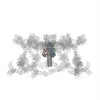
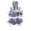
| Title | Fitting of the gp9 structure in the EM density of bacteriophage T4 extended tail | ||||||
 Components Components | Baseplate structural protein Gp9 | ||||||
 Keywords Keywords | VIRAL PROTEIN / Structural protein | ||||||
| Function / homology |  Function and homology information Function and homology informationvirus tail, baseplate / viral tail assembly / viral release from host cell Similarity search - Function | ||||||
| Biological species |  Enterobacteria phage T4 (virus) Enterobacteria phage T4 (virus) | ||||||
| Method | ELECTRON MICROSCOPY / single particle reconstruction / cryo EM / Resolution: 15 Å | ||||||
 Authors Authors | Kostyuchenko, V.A. | ||||||
 Citation Citation | Journal: Nat Struct Mol Biol / Year: 2005 Title: The tail structure of bacteriophage T4 and its mechanism of contraction. Authors: Victor A Kostyuchenko / Paul R Chipman / Petr G Leiman / Fumio Arisaka / Vadim V Mesyanzhinov / Michael G Rossmann /  Abstract: Bacteriophage T4 and related viruses have a contractile tail that serves as an efficient mechanical device for infecting bacteria. A three-dimensional cryo-EM reconstruction of the mature T4 tail ...Bacteriophage T4 and related viruses have a contractile tail that serves as an efficient mechanical device for infecting bacteria. A three-dimensional cryo-EM reconstruction of the mature T4 tail assembly at 15-A resolution shows the hexagonal dome-shaped baseplate, the extended contractile sheath, the long tail fibers attached to the baseplate and the collar formed by six whiskers that interact with the long tail fibers. Comparison with the structure of the contracted tail shows that tail contraction is associated with a substantial rearrangement of the domains within the sheath protein and results in shortening of the sheath to about one-third of its original length. During contraction, the tail tube extends beneath the baseplate by about one-half of its total length and rotates by 345 degrees , allowing it to cross the host's periplasmic space. | ||||||
| History |
|
- Structure visualization
| Movie |
Movie viewer |
|---|---|
| Structure viewer | Molecule: MolmilJmol/JSmol |
- Downloads & links
-Download
| PDBx/mmCIF format | 1zku.cif.gz | 925.5 KB | Display | PDBx/mmCIF format |
|---|---|---|---|---|
| PDB format | pdb1zku.ent.gz | 778.5 KB | Display | PDB format |
| PDBx/mmJSON format | 1zku.json.gz | Tree view | PDBx/mmJSON format | |
| Others |  Other downloads Other downloads |
-Validation report
| Arichive directory | https://data.pdbj.org/pub/pdb/validation_reports/zk/1zkuftp://data.pdbj.org/pub/pdb/validation_reports/zk/1zku | HTTPS FTP |
|---|
-Related structure data
| Related structure data |  1126MC 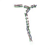 2bsgC M: map data used to model this data C: citing same article ( |
|---|---|
| Similar structure data |

-Links
 PDBj
PDBj- Assembly
| Deposited unit | 
|
|---|---|
| 1 |
|
-Components
| #1: Protein | Mass: 31024.725 Da / Num. of mol.: 18 Source method: isolated from a genetically manipulated source Source: (gene. exp.) Enterobacteria phage T4 (virus) / Genus: T4-like viruses / Species: Enterobacteria phage T4 sensu lato / Gene: 9 / References: UniProt: P10927#2: Chemical | ChemComp-EPE /   Mass: 238.305 Da / Num. of mol.: 18 / Source method: obtained synthetically / Formula: C8H18N2O4S / Comment: pH buffer*YM Mass: 238.305 Da / Num. of mol.: 18 / Source method: obtained synthetically / Formula: C8H18N2O4S / Comment: pH buffer*YM |
|---|
-Experimental details
-Experiment
| Experiment | Method: ELECTRON MICROSCOPY |
|---|---|
| EM experiment | Aggregation state: PARTICLE / 3D reconstruction method: single particle reconstruction |
- Sample preparation
| Component | Name: T4 / Type: VIRUS |
|---|---|
| Buffer solution | pH: 7 / Details: H2O |
| Specimen | Conc.: 10 mg/ml / Embedding applied: NO / Shadowing applied: NO / Staining applied: NO / Vitrification applied: YES |
| Specimen support | Details: COPPER GRID |
| Vitrification | Cryogen name: ETHANE / Details: LIQUID ETHANE |
- Electron microscopy imaging
| Microscopy | Model: FEI/PHILIPS CM300FEG/T / Date: Jan 10, 2003 |
|---|---|
| Electron gun | Electron source:  FIELD EMISSION GUN / Accelerating voltage: 300 kV / Illumination mode: SPOT SCAN FIELD EMISSION GUN / Accelerating voltage: 300 kV / Illumination mode: SPOT SCAN |
| Electron lens | Mode: BRIGHT FIELD / Nominal magnification: 45000 X / Calibrated magnification: 47000 X / Nominal defocus max: 3.2 nm / Nominal defocus min: 0.8 nm / Cs: 2 mm |
| Specimen holder | Temperature: 100 K |
| Image recording | Film or detector model: GENERIC FILM |
| Image scans | Num. digital images: 75 |
| Radiation | Protocol: SINGLE WAVELENGTH / Monochromatic (M) / Laue (L): M / Scattering type: x-ray |
| Radiation wavelength | Relative weight: 1 |
- Processing
| EM software |
| ||||||||||||
|---|---|---|---|---|---|---|---|---|---|---|---|---|---|
| CTF correction | Details: EACH IMAGE | ||||||||||||
| Symmetry | Point symmetry: C6 (6 fold cyclic) | ||||||||||||
| 3D reconstruction | Method: REAL SPACE SIRT / Resolution: 15 Å / Num. of particles: 3029 / Nominal pixel size: 3.97 Å / Actual pixel size: 3.97 Å / Magnification calibration: 47000 / Details: EM MAP DEPOSITED AS EMD-1126 / Symmetry type: POINT | ||||||||||||
| Atomic model building | Protocol: OTHER / Space: REAL Details: REFINEMENT PROTOCOL--MANUAL FITTING FOLLOWED BY REFINEMENT IN COLORES | ||||||||||||
| Atomic model building | PDB-ID: 1S2E Accession code: 1S2E / Source name: PDB / Type: experimental model | ||||||||||||
| Refinement | Highest resolution: 15 Å | ||||||||||||
| Refinement step | Cycle: LAST / Highest resolution: 15 Å
|
