 Movie
Movie Controller

[English] 日本語
 Yorodumi
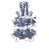
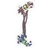
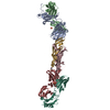
- PDB-1qex: BACTERIOPHAGE T4 GENE PRODUCT 9 (GP9), THE TRIGGER OF TAIL CONTRA... -
+ Open data

- Basic information
| Entry | Database: PDB / ID: 1qex | ||||||
|---|---|---|---|---|---|---|---|
| Title | BACTERIOPHAGE T4 GENE PRODUCT 9 (GP9), THE TRIGGER OF TAIL CONTRACTION AND THE LONG TAIL FIBERS CONNECTOR | ||||||
 Components Components | PROTEIN (BACTERIOPHAGE T4 GENE PRODUCT 9 (GP9)) | ||||||
 Keywords Keywords | VIRAL PROTEIN / BACTERIOPHAGE T4 / BASEPLATE / GENE PRODUCT 9 / OLIGOMERIZATION | ||||||
| Function / homology |  Function and homology information Function and homology informationvirus tail, baseplate / viral tail assembly / viral release from host cell Similarity search - Function | ||||||
| Biological species |  Enterobacteria phage T4 (virus) Enterobacteria phage T4 (virus) | ||||||
| Method |  X-RAY DIFFRACTION / MIRAS / Resolution: 2.3 Å X-RAY DIFFRACTION / MIRAS / Resolution: 2.3 Å | ||||||
 Authors Authors | Kostyuchenko, V.A. / Navruzbekov, G.A. / Kurochkina, L.P. / Strelkov, S.V. / Mesyanzhinov, V.V. / Rossmann, M.G. | ||||||
 Citation Citation | Journal: Structure Fold.Des. / Year: 1999 Title: The structure of bacteriophage T4 gene product 9: the trigger for tail contraction. Authors: Kostyuchenko, V.A. / Navruzbekov, G.A. / Kurochkina, L.P. / Strelkov, S.V. / Mesyanzhinov, V.V. / Rossmann, M.G. #1: Journal: J.Mol.Biol. / Year: 1993 Title: Crystallization and preliminary crystallographic characterization of bacteriophage T4 baseplate protein encoded by gene 9. Authors: Strelkov, S.V. / Zurabishvili, T.G. / Nepluev, I.V. / Efimov, V.P. / Isupov, M.N. / Harutyunyan, E.H. / Mesyanzhinov, V.V. | ||||||
| History |
|
- Structure visualization
| Structure viewer | Molecule: MolmilJmol/JSmol |
|---|
- Downloads & links
-Download
| PDBx/mmCIF format | 1qex.cif.gz | 125.5 KB | Display | PDBx/mmCIF format |
|---|---|---|---|---|
| PDB format | pdb1qex.ent.gz | 98.9 KB | Display | PDB format |
| PDBx/mmJSON format | 1qex.json.gz | Tree view | PDBx/mmJSON format | |
| Others |  Other downloads Other downloads |
-Validation report
| Arichive directory | https://data.pdbj.org/pub/pdb/validation_reports/qe/1qexftp://data.pdbj.org/pub/pdb/validation_reports/qe/1qex | HTTPS FTP |
|---|
-Related structure data

-Links
 PDBj
PDBj- Assembly
| Deposited unit | 
| ||||||||
|---|---|---|---|---|---|---|---|---|---|
| 1 | 
| ||||||||
| Unit cell |
| ||||||||
| Noncrystallographic symmetry (NCS) | NCS oper: (Code: given Matrix: (-0.49937, 0.86639, 0.00032), Vector: |
-Components
| #1: Protein | Mass: 31024.725 Da / Num. of mol.: 2 Source method: isolated from a genetically manipulated source Source: (gene. exp.) Enterobacteria phage T4 (virus) / Genus: T4-like viruses / Species: Enterobacteria phage T4 sensu lato / Gene: 9 / Plasmid: PET-23D / Species (production host): Escherichia coli / Production host:  #2: Chemical |   Mass: 238.305 Da / Num. of mol.: 2 / Source method: obtained synthetically / Formula: C8H18N2O4S / Comment: pH buffer*YM Mass: 238.305 Da / Num. of mol.: 2 / Source method: obtained synthetically / Formula: C8H18N2O4S / Comment: pH buffer*YM#3: Water | ChemComp-HOH / |  Mass: 18.015 Da / Num. of mol.: 292 / Source method: isolated from a natural source / Formula: H2O Mass: 18.015 Da / Num. of mol.: 292 / Source method: isolated from a natural source / Formula: H2O |
|---|
-Experimental details
-Experiment
| Experiment | Method: X-RAY DIFFRACTION / Number of used crystals: 1 |
|---|
- Sample preparation
| Crystal | Density Matthews: 3.1 Å3/Da / Density % sol: 60 % | ||||||||||||||||||||||||||||||
|---|---|---|---|---|---|---|---|---|---|---|---|---|---|---|---|---|---|---|---|---|---|---|---|---|---|---|---|---|---|---|---|
| Crystal grow | pH: 7.5 / Details: 28% PEG-400 0.2M CACL2 HEPES-NA BUFFER, PH 7.5 | ||||||||||||||||||||||||||||||
| Crystal | *PLUS | ||||||||||||||||||||||||||||||
| Crystal grow | *PLUS Method: vapor diffusion, hanging drop | ||||||||||||||||||||||||||||||
| Components of the solutions | *PLUS
|
-Data collection
| Diffraction | Mean temperature: 110 K |
|---|---|
| Diffraction source | Source: ROTATING ANODE / Type: RIGAKU RU200 / Wavelength: 1.5418 |
| Detector | Type: RIGAKU / Detector: IMAGE PLATE / Date: Jun 1, 1997 / Details: MIRRORS |
| Radiation | Monochromator: NI FILTER / Protocol: SINGLE WAVELENGTH / Monochromatic (M) / Laue (L): M / Scattering type: x-ray |
| Radiation wavelength | Wavelength: 1.5418 Å / Relative weight: 1 |
| Reflection | Resolution: 2.3→40 Å / Num. obs: 33400 / % possible obs: 97.5 % / Observed criterion σ(I): -2 / Redundancy: 5.8 % / Biso Wilson estimate: 31.7 Å2 / Rmerge(I) obs: 0.071 / Net I/σ(I): 15 |
| Reflection shell | Resolution: 2.3→2.4 Å / Redundancy: 5.6 % / Rmerge(I) obs: 0.17 / Mean I/σ(I) obs: 5 / % possible all: 96.3 |
| Reflection shell | *PLUS % possible obs: 96.3 % |
- Processing
| Software |
| ||||||||||||||||||||||||||||||||||||||||||||||||||||||||||||||||||||||||||||||||
|---|---|---|---|---|---|---|---|---|---|---|---|---|---|---|---|---|---|---|---|---|---|---|---|---|---|---|---|---|---|---|---|---|---|---|---|---|---|---|---|---|---|---|---|---|---|---|---|---|---|---|---|---|---|---|---|---|---|---|---|---|---|---|---|---|---|---|---|---|---|---|---|---|---|---|---|---|---|---|---|---|---|
| Refinement | Method to determine structure: MIRAS / Resolution: 2.3→20 Å / Rfactor Rfree error: 0.007 / Data cutoff high rms absF: 5466890.49 / Isotropic thermal model: GROUP / Cross valid method: THROUGHOUT / σ(F): 2 Details: DISORDERED REGIONS WERE MODELLED STEREOCHEMICALLY AND MARKED BY HIGH B-FACTORS
| ||||||||||||||||||||||||||||||||||||||||||||||||||||||||||||||||||||||||||||||||
| Solvent computation | Solvent model: FLAT MODEL / Bsol: 36.78 Å2 / ksol: 0.33 e/Å3 | ||||||||||||||||||||||||||||||||||||||||||||||||||||||||||||||||||||||||||||||||
| Displacement parameters | Biso mean: 34.5 Å2
| ||||||||||||||||||||||||||||||||||||||||||||||||||||||||||||||||||||||||||||||||
| Refine analyze |
| ||||||||||||||||||||||||||||||||||||||||||||||||||||||||||||||||||||||||||||||||
| Refinement step | Cycle: LAST / Resolution: 2.3→20 Å
| ||||||||||||||||||||||||||||||||||||||||||||||||||||||||||||||||||||||||||||||||
| Refine LS restraints |
| ||||||||||||||||||||||||||||||||||||||||||||||||||||||||||||||||||||||||||||||||
| LS refinement shell | Resolution: 2.3→2.4 Å / Rfactor Rfree error: 0.02 / Total num. of bins used: 8
| ||||||||||||||||||||||||||||||||||||||||||||||||||||||||||||||||||||||||||||||||
| Xplor file |
| ||||||||||||||||||||||||||||||||||||||||||||||||||||||||||||||||||||||||||||||||
| Software | *PLUS Name: CNS / Version: 0.5 / Classification: refinement | ||||||||||||||||||||||||||||||||||||||||||||||||||||||||||||||||||||||||||||||||
| Refinement | *PLUS Lowest resolution: 20 Å / σ(F): 2 / % reflection Rfree: 4.9 % / Rfactor obs: 0.23 | ||||||||||||||||||||||||||||||||||||||||||||||||||||||||||||||||||||||||||||||||
| Solvent computation | *PLUS | ||||||||||||||||||||||||||||||||||||||||||||||||||||||||||||||||||||||||||||||||
| Displacement parameters | *PLUS Biso mean: 34.5 Å2 | ||||||||||||||||||||||||||||||||||||||||||||||||||||||||||||||||||||||||||||||||
| Refine LS restraints | *PLUS
| ||||||||||||||||||||||||||||||||||||||||||||||||||||||||||||||||||||||||||||||||
| LS refinement shell | *PLUS Highest resolution: 2.3 Å / Rfactor Rfree: 0.304 / % reflection Rfree: 5.5 % / Rfactor Rwork: 0.28 |
