 Movie
Movie Controller

[English] 日本語
 Yorodumi
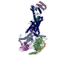
- PDB-2b5u: Crystal Structure Of Colicin E3 V206C Mutant In Complex With Its ... -
+ Open data

- Basic information
| Entry | Database: PDB / ID: 2b5u | ||||||
|---|---|---|---|---|---|---|---|
| Title | Crystal Structure Of Colicin E3 V206C Mutant In Complex With Its Immunity Protein | ||||||
 Components Components |
| ||||||
 Keywords Keywords | RIBOSOME INHIBITOR / HYDROLASE / High resolution crystal structure / Colicin E3 / Immunity Protein / Ribosome inactivation | ||||||
| Function / homology |  Function and homology information Function and homology informationextrachromosomal circular DNA / bacteriocin immunity / hydrolase activity, acting on ester bonds / toxic substance binding / ribosome binding / endonuclease activity / Lyases; Phosphorus-oxygen lyases / killing of cells of another organism / transmembrane transporter binding / tRNA binding ...extrachromosomal circular DNA / bacteriocin immunity / hydrolase activity, acting on ester bonds / toxic substance binding / ribosome binding / endonuclease activity / Lyases; Phosphorus-oxygen lyases / killing of cells of another organism / transmembrane transporter binding / tRNA binding / lyase activity / defense response to bacterium / rRNA binding / extracellular region Similarity search - Function | ||||||
| Biological species |  | ||||||
| Method |  X-RAY DIFFRACTION / SYNCHROTRON / FOURIER SYNTHESIS / Resolution: 2.3 Å X-RAY DIFFRACTION / SYNCHROTRON / FOURIER SYNTHESIS / Resolution: 2.3 Å | ||||||
 Authors Authors | Nallini Vijayarangan, A. / Nithianantham, S. / Nan, W. / Jakes, K. / Shoham, M. | ||||||
 Citation Citation | Journal: TO BE PUBLISHED Title: Crystal Structure Of Colicin E3 In Complex With Its Immunity Protein Authors: Nallini Vijayarangan, A. / Nithianantham, S. / Nan, W. / Jakes, K. / Shoham, M. | ||||||
| History |
|
- Structure visualization
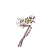
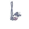
| Structure viewer | Molecule: MolmilJmol/JSmol |
|---|
- Downloads & links
-Download
| PDBx/mmCIF format | 2b5u.cif.gz | 225.1 KB | Display | PDBx/mmCIF format |
|---|---|---|---|---|
| PDB format | pdb2b5u.ent.gz | 179.9 KB | Display | PDB format |
| PDBx/mmJSON format | 2b5u.json.gz | Tree view | PDBx/mmJSON format | |
| Others |  Other downloads Other downloads |
-Validation report
| Arichive directory | https://data.pdbj.org/pub/pdb/validation_reports/b5/2b5uftp://data.pdbj.org/pub/pdb/validation_reports/b5/2b5u | HTTPS FTP |
|---|
-Related structure data
| Related structure data | 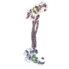 1jchS S: Starting model for refinement |
|---|---|
| Similar structure data |


-Links
 PDBj
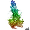
PDBj- Assembly
| Deposited unit | 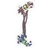
| ||||||||
|---|---|---|---|---|---|---|---|---|---|
| 1 | 
| ||||||||
| 2 | 
| ||||||||
| Unit cell |
|
-Components
| #1: Protein | Mass: 58047.023 Da / Num. of mol.: 2 / Mutation: V206C Source method: isolated from a genetically manipulated source Source: (gene. exp.) Production host: Strain (production host): W3110 References: UniProt: P00646, Hydrolases; Acting on ester bonds #2: Protein | Mass: 9779.565 Da / Num. of mol.: 2 Source method: isolated from a genetically manipulated source Source: (gene. exp.) Production host: Strain (production host): W3110 / References: UniProt: P02984 #3: Chemical |   Mass: 192.124 Da / Num. of mol.: 2 / Source method: obtained synthetically / Formula: C6H8O7 Mass: 192.124 Da / Num. of mol.: 2 / Source method: obtained synthetically / Formula: C6H8O7#4: Water | ChemComp-HOH / |  Mass: 18.015 Da / Num. of mol.: 219 / Source method: isolated from a natural source / Formula: H2O Mass: 18.015 Da / Num. of mol.: 219 / Source method: isolated from a natural source / Formula: H2O |
|---|
-Experimental details
-Experiment
| Experiment | Method: X-RAY DIFFRACTION / Number of used crystals: 1 |
|---|
- Sample preparation
| Crystal | Density Matthews: 3.92 Å3/Da / Density % sol: 66 % |
|---|---|
| Crystal grow | Temperature: 277 K / Method: vapor diffusion / pH: 5.6 Details: Sodium citrate, pH 5.6, VAPOR DIFFUSION, temperature 277K |
-Data collection
| Diffraction | Mean temperature: 100 K |
|---|---|
| Diffraction source | Source: SYNCHROTRON / Site: APS  / Beamline: 19-BM / Wavelength: 1.0332 Å / Beamline: 19-BM / Wavelength: 1.0332 Å |
| Detector | Detector: CCD / Date: Aug 3, 2005 / Details: Mirrors |
| Radiation | Monochromator: Si-111, Si-220 / Protocol: SINGLE WAVELENGTH / Monochromatic (M) / Laue (L): M / Scattering type: x-ray |
| Radiation wavelength | Wavelength: 1.0332 Å / Relative weight: 1 |
| Reflection | Resolution: 2.3→50 Å / Num. all: 88792 / Num. obs: 88240 / % possible obs: 99.4 % / Observed criterion σ(F): 0 / Observed criterion σ(I): 2 / Redundancy: 3.6 % / Rmerge(I) obs: 0.073 / Χ2: 1 |
| Reflection shell | Resolution: 2.3→2.38 Å / % possible obs: 97.7 % / Redundancy: 3.2 % / Rmerge(I) obs: 0.465 / Num. measured obs: 8682 / Χ2: 1.37 / % possible all: 97 |
- Processing
| Software |
| ||||||||||||||||||||||||||||
|---|---|---|---|---|---|---|---|---|---|---|---|---|---|---|---|---|---|---|---|---|---|---|---|---|---|---|---|---|---|
| Refinement | Method to determine structure: FOURIER SYNTHESIS Starting model: PDB ENTRY 1JCH Resolution: 2.3→44.4 Å / σ(F): 0 / Stereochemistry target values: Engh & Huber
| ||||||||||||||||||||||||||||
| Solvent computation | Bsol: 39.605 Å2 | ||||||||||||||||||||||||||||
| Displacement parameters | Biso mean: 51.725 Å2
| ||||||||||||||||||||||||||||
| Refinement step | Cycle: LAST / Resolution: 2.3→44.4 Å
| ||||||||||||||||||||||||||||
| Refine LS restraints |
| ||||||||||||||||||||||||||||
| Xplor file |
|
