Movie
Movie Controller

 Yorodumi
+ Open data

- Basic information
| Entry | Database: EMDB / ID: EMD-1126 | |||||||||
|---|---|---|---|---|---|---|---|---|---|---|
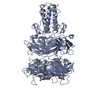
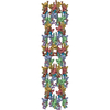
| Title | The tail structure of bacteriophage T4 and its mechanism of contraction. | |||||||||
 Map data Map data | EM density map for the extended tail of bacteriophage T4 | |||||||||
 Sample Sample |
| |||||||||
| Function / homology |  Function and homology information Function and homology informationvirus tail, sheath / symbiont genome ejection through host cell envelope, contractile tail mechanism / virus tail, baseplate / viral tail assembly / viral release from host cell / virion component Similarity search - Function | |||||||||
| Biological species |  Enterobacteria phage T4 (virus) Enterobacteria phage T4 (virus) | |||||||||
| Method | single particle reconstruction / cryo EM / negative staining / Resolution: 15.0 Å | |||||||||
 Authors Authors | Kostyuchenko VA / Chipman PR / Leiman PG / Arisaka F / Mesyanzhinov VV / Rossmann MG | |||||||||
 Citation Citation | Journal: Nat Struct Mol Biol / Year: 2005 Title: The tail structure of bacteriophage T4 and its mechanism of contraction. Authors: Victor A Kostyuchenko / Paul R Chipman / Petr G Leiman / Fumio Arisaka / Vadim V Mesyanzhinov / Michael G Rossmann /  Abstract: Bacteriophage T4 and related viruses have a contractile tail that serves as an efficient mechanical device for infecting bacteria. A three-dimensional cryo-EM reconstruction of the mature T4 tail ...Bacteriophage T4 and related viruses have a contractile tail that serves as an efficient mechanical device for infecting bacteria. A three-dimensional cryo-EM reconstruction of the mature T4 tail assembly at 15-A resolution shows the hexagonal dome-shaped baseplate, the extended contractile sheath, the long tail fibers attached to the baseplate and the collar formed by six whiskers that interact with the long tail fibers. Comparison with the structure of the contracted tail shows that tail contraction is associated with a substantial rearrangement of the domains within the sheath protein and results in shortening of the sheath to about one-third of its original length. During contraction, the tail tube extends beneath the baseplate by about one-half of its total length and rotates by 345 degrees , allowing it to cross the host's periplasmic space. | |||||||||
| History |
|
- Structure visualization
| Movie |
Movie viewer |
|---|---|
| Structure viewer | EM map: SurfViewMolmilJmol/JSmol |
| Supplemental images |

- Downloads & links
-EMDB archive
| Map data | emd_1126.map.gz | 18.4 MB | EMDB map data format | |
|---|---|---|---|---|
| Header (meta data) | emd-1126-v30.xmlemd-1126.xml | 9.2 KB 9.2 KB | Display Display | EMDB header |
| Images |  1126.gif 1126.gif | 11.6 KB | ||
| Archive directory |  http://ftp.pdbj.org/pub/emdb/structures/EMD-1126ftp://ftp.pdbj.org/pub/emdb/structures/EMD-1126 http://ftp.pdbj.org/pub/emdb/structures/EMD-1126ftp://ftp.pdbj.org/pub/emdb/structures/EMD-1126 | HTTPS FTP |
-Related structure data
| Related structure data | 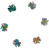 1zkuMC 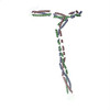 2bsgMC 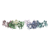 3fohM 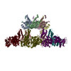 3j2mM M: atomic model generated by this map C: citing same article ( |
|---|---|
| Similar structure data |
-Links
| EMDB pages | EMDB (EBI/PDBe) / EMDataResource |
|---|---|
| Related items in Molecule of the Month |


-Map
| File | Download / File: emd_1126.map.gz / Format: CCP4 / Size: 45.9 MB / Type: IMAGE STORED AS FLOATING POINT NUMBER (4 BYTES) | ||||||||||||||||||||||||||||||||||||||||||||||||||||||||||||||||||||
|---|---|---|---|---|---|---|---|---|---|---|---|---|---|---|---|---|---|---|---|---|---|---|---|---|---|---|---|---|---|---|---|---|---|---|---|---|---|---|---|---|---|---|---|---|---|---|---|---|---|---|---|---|---|---|---|---|---|---|---|---|---|---|---|---|---|---|---|---|---|
| Annotation | EM density map for the extended tail of bacteriophage T4 | ||||||||||||||||||||||||||||||||||||||||||||||||||||||||||||||||||||
| Projections & slices | Image control
Images are generated by Spider. generated in cubic-lattice coordinate | ||||||||||||||||||||||||||||||||||||||||||||||||||||||||||||||||||||
| Voxel size | X=Y=Z: 3.97 Å | ||||||||||||||||||||||||||||||||||||||||||||||||||||||||||||||||||||
| Density |
| ||||||||||||||||||||||||||||||||||||||||||||||||||||||||||||||||||||
| Symmetry | Space group: 1 | ||||||||||||||||||||||||||||||||||||||||||||||||||||||||||||||||||||
| Details | EMDB XML:
CCP4 map header:
| ||||||||||||||||||||||||||||||||||||||||||||||||||||||||||||||||||||
 Z (Sec.)
 X (Row.)
 Y (Col.)


-Supplemental data
- Sample components
-Entire : native T4 phage
| Entire | Name: native T4 phage |
|---|---|
| Components |
|
-Supramolecule #1000: native T4 phage
| Supramolecule | Name: native T4 phage / type: sample / ID: 1000 / Oligomeric state: hexameric tails bound to 5-2 capsids / Number unique components: 1 |
|---|
-Supramolecule #1: Enterobacteria phage T4
| Supramolecule | Name: Enterobacteria phage T4 / type: virus / ID: 1 / Details: native phage / NCBI-ID: 10665 / Sci species name: Enterobacteria phage T4 / Virus type: VIRION / Virus isolate: STRAIN / Virus enveloped: No / Virus empty: No |
|---|---|
| Host (natural) | Organism:  |
-Experimental details
-Structure determination
| Method | negative staining, cryo EM |
|---|---|
 Processing Processing | single particle reconstruction |
| Aggregation state | particle |
-Sample preparation
| Buffer | pH: 7 / Details: H20 |
|---|---|
| Staining | Type: NEGATIVE / Details: cryo |
| Grid | Details: 200 mesh copper grids |
| Vitrification | Cryogen name: ETHANE |
- Electron microscopy
| Microscope | FEI/PHILIPS CM300FEG/ST |
|---|---|
| Date | Jan 10, 2003 |
| Image recording | Digitization - Scanner: ZEISS SCAI / Digitization - Sampling interval: 7 µm / Number real images: 89 / Bits/pixel: 12 |
| Electron beam | Acceleration voltage: 300 kV / Electron source:  FIELD EMISSION GUN FIELD EMISSION GUN |
| Electron optics | Calibrated magnification: 47000 / Illumination mode: SPOT SCAN / Imaging mode: BRIGHT FIELD / Cs: 2 mm / Nominal defocus max: 4.0 µm / Nominal defocus min: 1.0 µm / Nominal magnification: 45000 |
| Sample stage | Specimen holder model: GATAN LIQUID NITROGEN |
-Image processing
| CTF correction | Details: each particle image |
|---|---|
| Final reconstruction | Applied symmetry - Point group: C6 (6 fold cyclic) / Algorithm: OTHER / Resolution.type: BY AUTHOR / Resolution: 15.0 Å / Resolution method: FSC 0.5 CUT-OFF / Software - Name: SPIDER-like Details: reconstruction done with in-house implementation of BP RP algorithm from SPIDER to support non-cubicreconstruction volumes Number images used: 3029 |
-Atomic model buiding 1
| Initial model | PDB ID: |
|---|---|
| Refinement | Space: REAL |
| Output model | PDB-1zku: PDB-2bsg: PDB-3foh: PDB-3j2m: |

-Atomic model buiding 2
| Initial model | PDB ID: |
|---|---|
| Refinement | Space: REAL |
| Output model | PDB-1zku: PDB-2bsg: PDB-3foh: PDB-3j2m: |