 Movie
Movie Controller

[English] 日本語
 Yorodumi
- PDB-3foh: Fitting of gp18M crystal structure into 3D cryo-EM reconstruction... -
+ Open data

- Basic information
| Entry | Database: PDB / ID: 3foh | ||||||
|---|---|---|---|---|---|---|---|
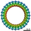
| Title | Fitting of gp18M crystal structure into 3D cryo-EM reconstruction of bacteriophage T4 extended tail | ||||||
 Components Components | Tail sheath protein Gp18 | ||||||
 Keywords Keywords | VIRAL PROTEIN / Alpha-beta / viral structural protein / bacteriophage T4 / tail sheath | ||||||
| Function / homology |  Function and homology information Function and homology informationvirus tail, sheath / symbiont genome ejection through host cell envelope, contractile tail mechanism Similarity search - Function | ||||||
| Biological species |  Enterobacteria phage T4 (virus) Enterobacteria phage T4 (virus) | ||||||
| Method | ELECTRON MICROSCOPY / single particle reconstruction / cryo EM / Resolution: 15 Å | ||||||
 Authors Authors | Aksyuk, A.A. / Leiman, P.G. / Kurochkina, L.P. / Shneider, M.M. / Kostyuchenko, V.A. / Mesyanzhinov, V.V. / Rossmann, M.G. | ||||||
 Citation Citation | Journal: EMBO J / Year: 2009 Title: The tail sheath structure of bacteriophage T4: a molecular machine for infecting bacteria. Authors: Anastasia A Aksyuk / Petr G Leiman / Lidia P Kurochkina / Mikhail M Shneider / Victor A Kostyuchenko / Vadim V Mesyanzhinov / Michael G Rossmann /  Abstract: The contractile tail of bacteriophage T4 is a molecular machine that facilitates very high viral infection efficiency. Its major component is a tail sheath, which contracts during infection to less ...The contractile tail of bacteriophage T4 is a molecular machine that facilitates very high viral infection efficiency. Its major component is a tail sheath, which contracts during infection to less than half of its initial length. The sheath consists of 138 copies of the tail sheath protein, gene product (gp) 18, which surrounds the central non-contractile tail tube. The contraction of the sheath drives the tail tube through the outer membrane, creating a channel for the viral genome delivery. A crystal structure of about three quarters of gp18 has been determined and was fitted into cryo-electron microscopy reconstructions of the tail sheath before and after contraction. It was shown that during contraction, gp18 subunits slide over each other with no apparent change in their structure. | ||||||
| History |
|
- Structure visualization
| Movie |
Movie viewer |
|---|---|
| Structure viewer | Molecule: MolmilJmol/JSmol |
- Downloads & links
-Download
| PDBx/mmCIF format | 3foh.cif.gz | 1 MB | Display | PDBx/mmCIF format |
|---|---|---|---|---|
| PDB format | pdb3foh.ent.gz | 894.2 KB | Display | PDB format |
| PDBx/mmJSON format | 3foh.json.gz | Tree view | PDBx/mmJSON format | |
| Others |  Other downloads Other downloads |
-Validation report
| Arichive directory | https://data.pdbj.org/pub/pdb/validation_reports/fo/3fohftp://data.pdbj.org/pub/pdb/validation_reports/fo/3foh | HTTPS FTP |
|---|
-Related structure data
| Related structure data |  1126M  3fo8C  3foaC 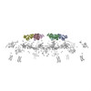 3foiC C: citing same article ( M: map data used to model this data |
|---|---|
| Similar structure data |
-Links
 PDBj
PDBj- Assembly
| Deposited unit | 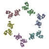
|
|---|---|
| 1 |
|
-Components
| #1: Protein | Mass: 54645.965 Da / Num. of mol.: 6 / Fragment: deletion mutant gp18M: UNP Residues 1-510 / Mutation: R510P Source method: isolated from a genetically manipulated source Source: (gene. exp.) Enterobacteria phage T4 (virus) / Gene: 18, GB AAA32541 / Plasmid: pET22 / Production host:  Sequence details | AUTHORS STATE THAT THE SEQUENCE COMPLETELY AGREES WITH THE GENBANK ENTRY AAA32541, PUBMED REPORT ...AUTHORS STATE THAT THE SEQUENCE COMPLETELY | |
|---|
-Experimental details
-Experiment
| Experiment | Method: ELECTRON MICROSCOPY |
|---|---|
| EM experiment | Aggregation state: PARTICLE / 3D reconstruction method: single particle reconstruction |
- Sample preparation
| Component |
| |||||||||||||||
|---|---|---|---|---|---|---|---|---|---|---|---|---|---|---|---|---|
| Buffer solution | pH: 7 / Details: H2O | |||||||||||||||
| Specimen | Embedding applied: NO / Shadowing applied: NO / Staining applied: NO / Vitrification applied: YES | |||||||||||||||
| Vitrification | Cryogen name: ETHANE |
- Electron microscopy imaging
| Microscopy | Model: FEI/PHILIPS CM300FEG/ST / Date: Jan 10, 2003 |
|---|---|
| Electron gun | Electron source:  FIELD EMISSION GUN / Accelerating voltage: 300 kV / Illumination mode: SPOT SCAN FIELD EMISSION GUN / Accelerating voltage: 300 kV / Illumination mode: SPOT SCAN |
| Electron lens | Mode: BRIGHT FIELD / Nominal magnification: 45000 X / Nominal defocus max: 4000 nm / Nominal defocus min: 1000 nm |
- Processing
| EM software |
| ||||||||||||
|---|---|---|---|---|---|---|---|---|---|---|---|---|---|
| Symmetry | Point symmetry: C6 (6 fold cyclic) | ||||||||||||
| 3D reconstruction | Resolution: 15 Å / Num. of particles: 3029 / Symmetry type: POINT | ||||||||||||
| Atomic model building | PDB-ID: 3FOA Accession code: 3FOA / Source name: PDB / Type: experimental model | ||||||||||||
| Refinement step | Cycle: LAST
|
