 Movie
Movie Controller

[English] 日本語
 Yorodumi
- PDB-3fo8: Crystal structure of the bacteriophage T4 tail sheath protein, pr... -
+ Open data

- Basic information
| Entry | Database: PDB / ID: 3fo8 | ||||||
|---|---|---|---|---|---|---|---|
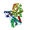
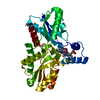
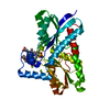
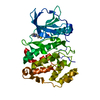
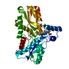
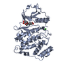
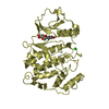
| Title | Crystal structure of the bacteriophage T4 tail sheath protein, protease resistant fragment gp18PR | ||||||
 Components Components | Tail sheath protein Gp18 | ||||||
 Keywords Keywords | VIRAL PROTEIN / mostly beta / viral structural protein / bacteriophage T4 / tail sheath | ||||||
| Function / homology |  Function and homology information Function and homology informationvirus tail, sheath / symbiont genome ejection through host cell envelope, contractile tail mechanism Similarity search - Function | ||||||
| Biological species |  Enterobacteria phage T4 (virus) Enterobacteria phage T4 (virus) | ||||||
| Method |  X-RAY DIFFRACTION / SYNCHROTRON / MAD / Resolution: 1.8 Å X-RAY DIFFRACTION / SYNCHROTRON / MAD / Resolution: 1.8 Å | ||||||
 Authors Authors | Aksyuk, A.A. / Leiman, P.G. / Kurochkina, L.P. / Shneider, M.M. / Kostyuchenko, V.A. / Mesyanzhinov, V.V. / Rossmann, M.G. | ||||||
 Citation Citation | Journal: EMBO J / Year: 2009 Title: The tail sheath structure of bacteriophage T4: a molecular machine for infecting bacteria. Authors: Anastasia A Aksyuk / Petr G Leiman / Lidia P Kurochkina / Mikhail M Shneider / Victor A Kostyuchenko / Vadim V Mesyanzhinov / Michael G Rossmann /  Abstract: The contractile tail of bacteriophage T4 is a molecular machine that facilitates very high viral infection efficiency. Its major component is a tail sheath, which contracts during infection to less ...The contractile tail of bacteriophage T4 is a molecular machine that facilitates very high viral infection efficiency. Its major component is a tail sheath, which contracts during infection to less than half of its initial length. The sheath consists of 138 copies of the tail sheath protein, gene product (gp) 18, which surrounds the central non-contractile tail tube. The contraction of the sheath drives the tail tube through the outer membrane, creating a channel for the viral genome delivery. A crystal structure of about three quarters of gp18 has been determined and was fitted into cryo-electron microscopy reconstructions of the tail sheath before and after contraction. It was shown that during contraction, gp18 subunits slide over each other with no apparent change in their structure. | ||||||
| History |
|
- Structure visualization
| Structure viewer | Molecule: MolmilJmol/JSmol |
|---|
- Downloads & links
-Download
| PDBx/mmCIF format | 3fo8.cif.gz | 75.7 KB | Display | PDBx/mmCIF format |
|---|---|---|---|---|
| PDB format | pdb3fo8.ent.gz | 57.5 KB | Display | PDB format |
| PDBx/mmJSON format | 3fo8.json.gz | Tree view | PDBx/mmJSON format | |
| Others |  Other downloads Other downloads |
-Validation report
| Arichive directory | https://data.pdbj.org/pub/pdb/validation_reports/fo/3fo8ftp://data.pdbj.org/pub/pdb/validation_reports/fo/3fo8 | HTTPS FTP |
|---|
-Related structure data


-Links
 PDBj
PDBj- Assembly
| Deposited unit | 
| |||||||||
|---|---|---|---|---|---|---|---|---|---|---|
| 1 |
| |||||||||
| 2 | x 24
| |||||||||
| Unit cell |
| |||||||||
| Components on special symmetry positions |
|
-Components
| #1: Protein | Mass: 29887.404 Da / Num. of mol.: 1 Fragment: protease resistant fragment gp18PR: UNP Residues 83-365 Source method: isolated from a genetically manipulated source Source: (gene. exp.) Enterobacteria phage T4 (virus) / Gene: 18, GB AAA32541.1 / Plasmid: pET22 / Production host:  |
|---|---|
| #2: Chemical | ChemComp-ACT /   Mass: 59.044 Da / Num. of mol.: 1 / Source method: obtained synthetically / Formula: C2H3O2 Mass: 59.044 Da / Num. of mol.: 1 / Source method: obtained synthetically / Formula: C2H3O2 |
| #3: Water | ChemComp-HOH /  Mass: 18.015 Da / Num. of mol.: 422 / Source method: isolated from a natural source / Formula: H2O Mass: 18.015 Da / Num. of mol.: 422 / Source method: isolated from a natural source / Formula: H2O |
| Sequence details | AUTHORS STATE THAT THE SEQUENCE COMPLETELY AGREES WITH THE GENBANK ENTRY AAA32541, PUBMED REPORT ...AUTHORS STATE THAT THE SEQUENCE COMPLETELY |
-Experimental details
-Experiment
| Experiment | Method: X-RAY DIFFRACTION / Number of used crystals: 1 |
|---|
- Sample preparation
| Crystal | Density Matthews: 2.94 Å3/Da / Density % sol: 58.12 % Description: The structure factor file contains Friedel pairs |
|---|---|
| Crystal grow | Temperature: 293 K / Method: vapor diffusion, hanging drop / pH: 8 Details: 1.2M Sodium acetate, 1M Imidazole pH 8.0, VAPOR DIFFUSION, HANGING DROP, temperature 293K |
-Data collection
| Diffraction | Mean temperature: 100 K | ||||||||||||
|---|---|---|---|---|---|---|---|---|---|---|---|---|---|
| Diffraction source | Source: SYNCHROTRON / Site: APS / Beamline: 23-ID-D / Wavelength: 0.97942, 0.97956, 0.97818 | ||||||||||||
| Detector | Type: MARMOSAIC 300 mm CCD / Detector: CCD / Date: Oct 10, 2007 / Details: Adjustable focusing mirrors in K-B geometry | ||||||||||||
| Radiation | Monochromator: Double crystal cryo-cooled Si(111) / Protocol: MAD / Scattering type: x-ray | ||||||||||||
| Radiation wavelength |
| ||||||||||||
| Reflection | Resolution: 1.8→36 Å / Num. obs: 61873 / % possible obs: 88.11 % / Observed criterion σ(I): 2 / Redundancy: 5.5 % / Rmerge(I) obs: 0.064 / Χ2: 2.867 / Net I/σ(I): 26.802 | ||||||||||||
| Reflection shell | Resolution: 1.8→1.84 Å / Redundancy: 4.4 % / Rmerge(I) obs: 0.794 / Mean I/σ(I) obs: 2 / Num. unique all: 2112 / Χ2: 1.19 / % possible all: 95.5 |
- Processing
| Software |
| |||||||||||||||||||||||||||||||||||||||||||||||||||||||||||||||||||||||||||||||||||||||||||||||||||||||||||||||||||||||||||||||||||||||||||||||||||
|---|---|---|---|---|---|---|---|---|---|---|---|---|---|---|---|---|---|---|---|---|---|---|---|---|---|---|---|---|---|---|---|---|---|---|---|---|---|---|---|---|---|---|---|---|---|---|---|---|---|---|---|---|---|---|---|---|---|---|---|---|---|---|---|---|---|---|---|---|---|---|---|---|---|---|---|---|---|---|---|---|---|---|---|---|---|---|---|---|---|---|---|---|---|---|---|---|---|---|---|---|---|---|---|---|---|---|---|---|---|---|---|---|---|---|---|---|---|---|---|---|---|---|---|---|---|---|---|---|---|---|---|---|---|---|---|---|---|---|---|---|---|---|---|---|---|---|---|---|
| Refinement | Method to determine structure: MAD / Resolution: 1.8→36 Å / Occupancy max: 1 / Occupancy min: 0.38 / FOM work R set: 0.835 / SU ML: 0.22 / σ(F): 1.88 / Stereochemistry target values: MAXIMUM LIKELIHOOD / Details: The Friedel pairs were used in phasing
| |||||||||||||||||||||||||||||||||||||||||||||||||||||||||||||||||||||||||||||||||||||||||||||||||||||||||||||||||||||||||||||||||||||||||||||||||||
| Solvent computation | Shrinkage radii: 0.9 Å / VDW probe radii: 1.11 Å / Solvent model: FLAT BULK SOLVENT MODEL / Bsol: 43.779 Å2 / ksol: 0.364 e/Å3 | |||||||||||||||||||||||||||||||||||||||||||||||||||||||||||||||||||||||||||||||||||||||||||||||||||||||||||||||||||||||||||||||||||||||||||||||||||
| Displacement parameters | Biso max: 63.35 Å2 / Biso mean: 22.108 Å2 / Biso min: 8.36 Å2
| |||||||||||||||||||||||||||||||||||||||||||||||||||||||||||||||||||||||||||||||||||||||||||||||||||||||||||||||||||||||||||||||||||||||||||||||||||
| Refinement step | Cycle: LAST / Resolution: 1.8→36 Å
| |||||||||||||||||||||||||||||||||||||||||||||||||||||||||||||||||||||||||||||||||||||||||||||||||||||||||||||||||||||||||||||||||||||||||||||||||||
| Refine LS restraints |
| |||||||||||||||||||||||||||||||||||||||||||||||||||||||||||||||||||||||||||||||||||||||||||||||||||||||||||||||||||||||||||||||||||||||||||||||||||
| LS refinement shell | Refine-ID: X-RAY DIFFRACTION / Total num. of bins used: 20
|
