 Movie
Movie Controller

[English] 日本語
 Yorodumi
- PDB-2q3j: Crystal structure of the His183Ala variant of Bacillus subtilis f... -
+ Open data

- Basic information
| Entry | Database: PDB / ID: 2q3j | ||||||
|---|---|---|---|---|---|---|---|
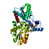
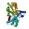
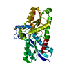
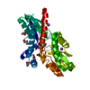
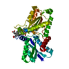
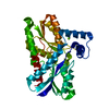
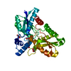
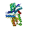
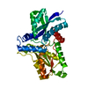
| Title | Crystal structure of the His183Ala variant of Bacillus subtilis ferrochelatase in complex with N-Methyl Mesoporphyrin | ||||||
 Components Components | Ferrochelatase | ||||||
 Keywords Keywords | LYASE / ROSSMANN FOLD / PI-HELIX / N-METHYL MESOPORPHYRIN IX / N-MeMP | ||||||
| Function / homology |  Function and homology information Function and homology informationcoproporphyrin ferrochelatase / protoporphyrin ferrochelatase activity / heme biosynthetic process / metal ion binding / cytoplasm Similarity search - Function | ||||||
| Biological species |  | ||||||
| Method |  X-RAY DIFFRACTION / SYNCHROTRON / MOLECULAR REPLACEMENT / Resolution: 2.39 Å X-RAY DIFFRACTION / SYNCHROTRON / MOLECULAR REPLACEMENT / Resolution: 2.39 Å | ||||||
 Authors Authors | Karlberg, T. / Thorvaldsen, O.H. / Al-Karadaghi, S. | ||||||
 Citation Citation | Journal: J.Mol.Biol. / Year: 2008 Title: Porphyrin binding and distortion and substrate specificity in the ferrochelatase reaction: the role of active site residues Authors: Karlberg, T. / Hansson, M.D. / Yengo, R.K. / Johansson, R. / Thorvaldsen, O.H. / Ferreira, G.C. / Hansson, M. / Al-Karadaghi, S. | ||||||
| History |
|
- Structure visualization
| Structure viewer | Molecule: MolmilJmol/JSmol |
|---|
- Downloads & links
-Download
| PDBx/mmCIF format | 2q3j.cif.gz | 79.8 KB | Display | PDBx/mmCIF format |
|---|---|---|---|---|
| PDB format | pdb2q3j.ent.gz | 57.7 KB | Display | PDB format |
| PDBx/mmJSON format | 2q3j.json.gz | Tree view | PDBx/mmJSON format | |
| Others |  Other downloads Other downloads |
-Validation report
| Arichive directory | https://data.pdbj.org/pub/pdb/validation_reports/q3/2q3jftp://data.pdbj.org/pub/pdb/validation_reports/q3/2q3j | HTTPS FTP |
|---|
-Related structure data
| Related structure data | 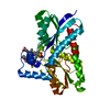 2q2nC  2q2oC  2h1wS C: citing same article ( S: Starting model for refinement |
|---|---|
| Similar structure data |

-Links
 PDBj
PDBj- Assembly
| Deposited unit | 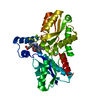
| ||||||||
|---|---|---|---|---|---|---|---|---|---|
| 1 |
| ||||||||
| Unit cell |
| ||||||||
| Details | The biological assembly is a monomer |
-Components
| #1: Protein | Mass: 35191.441 Da / Num. of mol.: 1 / Mutation: H183A Source method: isolated from a genetically manipulated source Source: (gene. exp.) |
|---|---|
| #2: Chemical | ChemComp-MG /   Mass: 24.305 Da / Num. of mol.: 1 / Source method: obtained synthetically / Formula: Mg Mass: 24.305 Da / Num. of mol.: 1 / Source method: obtained synthetically / Formula: Mg |
| #3: Chemical | ChemComp-H02 /   Mass: 582.732 Da / Num. of mol.: 1 / Source method: obtained synthetically / Formula: C35H42N4O4 Mass: 582.732 Da / Num. of mol.: 1 / Source method: obtained synthetically / Formula: C35H42N4O4 |
| #4: Water | ChemComp-HOH /  Mass: 18.015 Da / Num. of mol.: 146 / Source method: isolated from a natural source / Formula: H2O Mass: 18.015 Da / Num. of mol.: 146 / Source method: isolated from a natural source / Formula: H2O |
-Experimental details
-Experiment
| Experiment | Method: X-RAY DIFFRACTION / Number of used crystals: 1 |
|---|
- Sample preparation
| Crystal | Density Matthews: 2.03 Å3/Da / Density % sol: 39.27 % |
|---|---|
| Crystal grow | Temperature: 298 K / Method: vapor diffusion, hanging drop / pH: 7.4 Details: 25-30 % PEG 2000, 0.2 M MgCl2, 0.1 M TRIS-HCL, pH 7.4, VAPOR DIFFUSION, HANGING DROP, temperature 298K |
-Data collection
| Diffraction | Mean temperature: 100 K |
|---|---|
| Diffraction source | Source: SYNCHROTRON / Site: MAX II  / Beamline: I911-5 / Wavelength: 0.907 / Beamline: I911-5 / Wavelength: 0.907 |
| Detector | Type: MARMOSAIC 225 mm CCD / Detector: CCD / Date: Dec 7, 2005 |
| Radiation | Monochromator: DOUBLE CRYSTAL MONOCHROMATOR, SI(111) / Protocol: SINGLE WAVELENGTH / Monochromatic (M) / Laue (L): M / Scattering type: x-ray |
| Radiation wavelength | Wavelength: 0.907 Å / Relative weight: 1 |
| Reflection | Resolution: 2.39→30 Å / Num. all: 11903 / Num. obs: 11775 / % possible obs: 98.7 % / Observed criterion σ(I): 0 / Redundancy: 5.1 % / Rmerge(I) obs: 0.103 / Rsym value: 0.075 / Net I/σ(I): 18.4 |
| Reflection shell | Resolution: 2.39→2.53 Å / Redundancy: 5 % / Rmerge(I) obs: 0.299 / Mean I/σ(I) obs: 10.4 / Num. unique all: 1747 / Rsym value: 0.194 / % possible all: 94 |
- Processing
| Software |
| ||||||||||||||||||||||||||||
|---|---|---|---|---|---|---|---|---|---|---|---|---|---|---|---|---|---|---|---|---|---|---|---|---|---|---|---|---|---|
| Refinement | Method to determine structure: MOLECULAR REPLACEMENT Starting model: 2H1W Resolution: 2.39→20 Å / Cross valid method: THROUGHOUT / Stereochemistry target values: Engh & Huber
| ||||||||||||||||||||||||||||
| Refinement step | Cycle: LAST / Resolution: 2.39→20 Å
| ||||||||||||||||||||||||||||
| Refine LS restraints |
|
