 Movie
Movie Controller


+ Open data

- Basic information
| Entry | Database: PDB / ID: 6rwv | ||||||
|---|---|---|---|---|---|---|---|
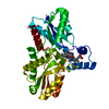
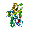
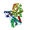
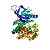
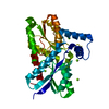
| Title | Structure of apo-LmCpfC | ||||||
 Components Components | Ferrochelatase | ||||||
 Keywords Keywords | METAL BINDING PROTEIN / Ferrochelatase / Prokaryotic heme biosynthesis / ferredoxin-like fold | ||||||
| Function / homology |  Function and homology information Function and homology informationcoproporphyrin ferrochelatase / protoporphyrin ferrochelatase activity / heme biosynthetic process / metal ion binding / cytoplasm Similarity search - Function | ||||||
| Biological species |  Listeria monocytogenes (bacteria) Listeria monocytogenes (bacteria) | ||||||
| Method |  X-RAY DIFFRACTION / SYNCHROTRON / MOLECULAR REPLACEMENT / Resolution: 1.63863795352 Å X-RAY DIFFRACTION / SYNCHROTRON / MOLECULAR REPLACEMENT / Resolution: 1.63863795352 Å | ||||||
 Authors Authors | Hofbauer, S. / Helm, J. / Djinovic-Carugo, K. / Furtmueller, P.G. | ||||||
| Funding support |  Austria, 1items Austria, 1items
| ||||||
 Citation Citation | Journal: Febs J. / Year: 2020 Title: Crystal structures and calorimetry reveal catalytically relevant binding mode of coproporphyrin and coproheme in coproporphyrin ferrochelatase. Authors: Hofbauer, S. / Helm, J. / Obinger, C. / Djinovic-Carugo, K. / Furtmuller, P.G. | ||||||
| History |
|
- Structure visualization
| Structure viewer | Molecule: MolmilJmol/JSmol |
|---|
- Downloads & links
-Download
| PDBx/mmCIF format | 6rwv.cif.gz | 174.6 KB | Display | PDBx/mmCIF format |
|---|---|---|---|---|
| PDB format | pdb6rwv.ent.gz | 114.6 KB | Display | PDB format |
| PDBx/mmJSON format | 6rwv.json.gz | Tree view | PDBx/mmJSON format | |
| Others |  Other downloads Other downloads |
-Validation report
| Arichive directory | https://data.pdbj.org/pub/pdb/validation_reports/rw/6rwvftp://data.pdbj.org/pub/pdb/validation_reports/rw/6rwv | HTTPS FTP |
|---|
-Related structure data
| Related structure data |  6sv3C  2hk6S S: Starting model for refinement C: citing same article ( |
|---|---|
| Similar structure data |


-Links
 PDBj
PDBj- Assembly
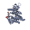
| Deposited unit | 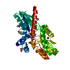
| ||||||||||||
|---|---|---|---|---|---|---|---|---|---|---|---|---|---|
| 1 |
| ||||||||||||
| Unit cell |
|
-Components
| #1: Protein | Mass: 35770.219 Da / Num. of mol.: 1 Source method: isolated from a genetically manipulated source Source: (gene. exp.) Listeria monocytogenes (bacteria)Gene: hemH, A4G43_07995, AF115_13645, AP101_13400, AP103_13395, AP112_12715, AP127_13135, AP130_13225, APD66_13050, ARS86_05675, B1N21_11380, B4Y57_13635, B5G78_12175, B5H07_07285, BRS71_03785, D3X95_ ...Gene: hemH, A4G43_07995, AF115_13645, AP101_13400, AP103_13395, AP112_12715, AP127_13135, AP130_13225, APD66_13050, ARS86_05675, B1N21_11380, B4Y57_13635, B5G78_12175, B5H07_07285, BRS71_03785, D3X95_05200, D3Y03_05130, D8K64_06195, EAJ22_07595, EAX63_13360, EFX44_12300, SG10_07760 Production host: References: UniProt: A0A3T2BSC5, UniProt: Q8Y565*PLUS, EC: 4.99.1.1 | ||||||
|---|---|---|---|---|---|---|---|
| #2: Chemical | ChemComp-GOL /   Mass: 92.094 Da / Num. of mol.: 7 / Source method: obtained synthetically / Formula: C3H8O3 Mass: 92.094 Da / Num. of mol.: 7 / Source method: obtained synthetically / Formula: C3H8O3#3: Chemical |   Mass: 94.971 Da / Num. of mol.: 2 / Source method: obtained synthetically / Formula: PO4 Mass: 94.971 Da / Num. of mol.: 2 / Source method: obtained synthetically / Formula: PO4#4: Chemical |   Mass: 39.098 Da / Num. of mol.: 2 / Source method: obtained synthetically / Formula: K Mass: 39.098 Da / Num. of mol.: 2 / Source method: obtained synthetically / Formula: K#5: Water | ChemComp-HOH / |  Mass: 18.015 Da / Num. of mol.: 268 / Source method: isolated from a natural source / Formula: H2O Mass: 18.015 Da / Num. of mol.: 268 / Source method: isolated from a natural source / Formula: H2O |
-Experimental details
-Experiment
| Experiment | Method: X-RAY DIFFRACTION / Number of used crystals: 1 |
|---|
- Sample preparation
| Crystal | Density Matthews: 2.61 Å3/Da / Density % sol: 52.91 % |
|---|---|
| Crystal grow | Temperature: 298 K / Method: vapor diffusion, sitting drop / Details: 16% w/v PEG 8000, 20% w/v Glycerol, 0.04 M KH2PO4 |
-Data collection
| Diffraction | Mean temperature: 100 K / Serial crystal experiment: N |
|---|---|
| Diffraction source | Source: SYNCHROTRON / Site: Diamond  / Beamline: I03 / Wavelength: 0.98 Å / Beamline: I03 / Wavelength: 0.98 Å |
| Detector | Type: DECTRIS EIGER X 16M / Detector: PIXEL / Date: Jan 24, 2019 |
| Radiation | Protocol: SINGLE WAVELENGTH / Monochromatic (M) / Laue (L): M / Scattering type: x-ray |
| Radiation wavelength | Wavelength: 0.98 Å / Relative weight: 1 |
| Reflection | Resolution: 1.63863795352→50.0905681247 Å / Num. obs: 44834 / % possible obs: 99.84 % / Redundancy: 2 % / Biso Wilson estimate: 18.060682719 Å2 / CC1/2: 0.997 / Rmerge(I) obs: 0.06407 / Rpim(I) all: 0.06407 / Rrim(I) all: 0.09061 / Net I/σ(I): 6.5 |
| Reflection shell | Resolution: 1.639→1.698 Å / Redundancy: 2 % / Rmerge(I) obs: 0.6822 / Mean I/σ(I) obs: 1.09 / Num. unique obs: 4416 / CC1/2: 0.435 / Rpim(I) all: 0.6822 / Rrim(I) all: 0.9648 / % possible all: 99.1 |
- Processing
| Software |
| |||||||||||||||||||||||||||||||||||||||||||||||||||||||||||||||||||||||||||||||||||||||||||||||||||||||||||||||||||||||
|---|---|---|---|---|---|---|---|---|---|---|---|---|---|---|---|---|---|---|---|---|---|---|---|---|---|---|---|---|---|---|---|---|---|---|---|---|---|---|---|---|---|---|---|---|---|---|---|---|---|---|---|---|---|---|---|---|---|---|---|---|---|---|---|---|---|---|---|---|---|---|---|---|---|---|---|---|---|---|---|---|---|---|---|---|---|---|---|---|---|---|---|---|---|---|---|---|---|---|---|---|---|---|---|---|---|---|---|---|---|---|---|---|---|---|---|---|---|---|---|---|
| Refinement | Method to determine structure: MOLECULAR REPLACEMENT Starting model: 2HK6 Resolution: 1.63863795352→50.0905681247 Å / SU ML: 0.19115048118 / Cross valid method: FREE R-VALUE / σ(F): 1.33645276556 / Phase error: 21.9177428424
| |||||||||||||||||||||||||||||||||||||||||||||||||||||||||||||||||||||||||||||||||||||||||||||||||||||||||||||||||||||||
| Solvent computation | Shrinkage radii: 0.9 Å / VDW probe radii: 1.11 Å | |||||||||||||||||||||||||||||||||||||||||||||||||||||||||||||||||||||||||||||||||||||||||||||||||||||||||||||||||||||||
| Displacement parameters | Biso mean: 22.3643472954 Å2 | |||||||||||||||||||||||||||||||||||||||||||||||||||||||||||||||||||||||||||||||||||||||||||||||||||||||||||||||||||||||
| Refinement step | Cycle: LAST / Resolution: 1.63863795352→50.0905681247 Å
| |||||||||||||||||||||||||||||||||||||||||||||||||||||||||||||||||||||||||||||||||||||||||||||||||||||||||||||||||||||||
| Refine LS restraints |
| |||||||||||||||||||||||||||||||||||||||||||||||||||||||||||||||||||||||||||||||||||||||||||||||||||||||||||||||||||||||
| LS refinement shell |
| |||||||||||||||||||||||||||||||||||||||||||||||||||||||||||||||||||||||||||||||||||||||||||||||||||||||||||||||||||||||
| Refinement TLS params. | Method: refined / Origin x: 15.3388808755 Å / Origin y: 41.8680056253 Å / Origin z: 31.9168207735 Å
| |||||||||||||||||||||||||||||||||||||||||||||||||||||||||||||||||||||||||||||||||||||||||||||||||||||||||||||||||||||||
| Refinement TLS group | Selection details: (chain A and resseq 3:312) |
