 Movie
Movie Controller

[English] 日本語
 Yorodumi
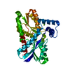
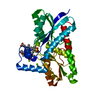
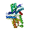
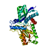
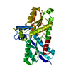
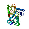
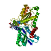
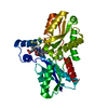
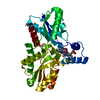
- PDB-2hk6: Crystal Structure of B. subtilis ferrochelatase with Iron bound a... -
+ Open data

- Basic information
| Entry | Database: PDB / ID: 2hk6 | ||||||
|---|---|---|---|---|---|---|---|
| Title | Crystal Structure of B. subtilis ferrochelatase with Iron bound at the active site | ||||||
 Components Components | Ferrochelatase | ||||||
 Keywords Keywords | LYASE / Heme biosynthesis / Iron / Metal-binding / Porphyrin biosynthesis | ||||||
| Function / homology |  Function and homology information Function and homology informationcoproporphyrin ferrochelatase / protoporphyrin ferrochelatase activity / heme biosynthetic process / metal ion binding / cytoplasm Similarity search - Function | ||||||
| Biological species |  | ||||||
| Method |  X-RAY DIFFRACTION / SYNCHROTRON / MOLECULAR REPLACEMENT / Resolution: 1.71 Å X-RAY DIFFRACTION / SYNCHROTRON / MOLECULAR REPLACEMENT / Resolution: 1.71 Å | ||||||
 Authors Authors | Al-Karadaghi, S. / Karlberg, T. | ||||||
 Citation Citation | Journal: Biochemistry / Year: 2007 Title: Amino Acid Residues His183 and Glu264 in Bacillus subtilis Ferrochelatase Direct and Facilitate the Insertion of Metal Ion into Protoporphyrin IX Authors: Hansson, M.D. / Karlberg, T. / Rahardja, M.A. / Al-Karadaghi, S. / Hansson, M. | ||||||
| History |
| ||||||
| Remark 600 | Heterogen The authors state that the charge state of FE is unknown. |
- Structure visualization
| Structure viewer | Molecule: MolmilJmol/JSmol |
|---|
- Downloads & links
-Download
| PDBx/mmCIF format | 2hk6.cif.gz | 86.1 KB | Display | PDBx/mmCIF format |
|---|---|---|---|---|
| PDB format | pdb2hk6.ent.gz | 63.2 KB | Display | PDB format |
| PDBx/mmJSON format | 2hk6.json.gz | Tree view | PDBx/mmJSON format | |
| Others |  Other downloads Other downloads |
-Validation report
| Arichive directory | https://data.pdbj.org/pub/pdb/validation_reports/hk/2hk6ftp://data.pdbj.org/pub/pdb/validation_reports/hk/2hk6 | HTTPS FTP |
|---|
-Related structure data
| Related structure data |  2h1vC  2h1wC  1dozS S: Starting model for refinement C: citing same article ( |
|---|---|
| Similar structure data |

-Links
 PDBj
PDBj- Assembly
| Deposited unit | 
| ||||||||
|---|---|---|---|---|---|---|---|---|---|
| 1 |
| ||||||||
| Unit cell |
|
-Components
| #1: Protein | Mass: 35389.707 Da / Num. of mol.: 1 Source method: isolated from a genetically manipulated source Source: (gene. exp.) | ||||
|---|---|---|---|---|---|
| #2: Chemical | ChemComp-FE /   Mass: 55.845 Da / Num. of mol.: 4 / Source method: obtained synthetically / Formula: Fe Mass: 55.845 Da / Num. of mol.: 4 / Source method: obtained synthetically / Formula: Fe#3: Chemical |   Mass: 24.305 Da / Num. of mol.: 3 / Source method: obtained synthetically / Formula: Mg Mass: 24.305 Da / Num. of mol.: 3 / Source method: obtained synthetically / Formula: Mg#4: Water | ChemComp-HOH / |  Mass: 18.015 Da / Num. of mol.: 385 / Source method: isolated from a natural source / Formula: H2O Mass: 18.015 Da / Num. of mol.: 385 / Source method: isolated from a natural source / Formula: H2O |
-Experimental details
-Experiment
| Experiment | Method: X-RAY DIFFRACTION / Number of used crystals: 1 |
|---|
- Sample preparation
| Crystal | Density Matthews: 2.04 Å3/Da / Density % sol: 39.72 % |
|---|---|
| Crystal grow | Temperature: 288 K / Method: vapor diffusion / pH: 8 Details: 25% PEG 2000, 0.2 M MgCl2, 0.1 M Tris-HCl , pH 8.0, VAPOR DIFFUSION, temperature 288K |
-Data collection
| Diffraction | Mean temperature: 100 K |
|---|---|
| Diffraction source | Source: SYNCHROTRON / Site: MAX II  / Beamline: I911-3 / Wavelength: 1.0605 Å / Beamline: I911-3 / Wavelength: 1.0605 Å |
| Detector | Type: MAR CCD 165 mm / Detector: CCD / Date: Jun 8, 2005 |
| Radiation | Monochromator: Double crystal monochromator, Si(111) / Protocol: SINGLE WAVELENGTH / Monochromatic (M) / Laue (L): M / Scattering type: x-ray |
| Radiation wavelength | Wavelength: 1.0605 Å / Relative weight: 1 |
| Reflection | Resolution: 1.7→20 Å / Num. obs: 31884 / % possible obs: 100 % / Observed criterion σ(F): 0 / Observed criterion σ(I): 0 / Redundancy: 11.3 % / Rmerge(I) obs: 0.027 / Rsym value: 0.055 |
| Reflection shell | Resolution: 1.7→1.81 Å / % possible all: 95.6 |
- Processing
| Software |
| |||||||||||||||||||||||||||||||||||||||||||||||||||||||||||||||||||||||||||||||||||||||||||||||
|---|---|---|---|---|---|---|---|---|---|---|---|---|---|---|---|---|---|---|---|---|---|---|---|---|---|---|---|---|---|---|---|---|---|---|---|---|---|---|---|---|---|---|---|---|---|---|---|---|---|---|---|---|---|---|---|---|---|---|---|---|---|---|---|---|---|---|---|---|---|---|---|---|---|---|---|---|---|---|---|---|---|---|---|---|---|---|---|---|---|---|---|---|---|---|---|---|
| Refinement | Method to determine structure: MOLECULAR REPLACEMENT Starting model: PDB ENTRY 1DOZ Resolution: 1.71→19.64 Å / Cor.coef. Fo:Fc: 0.95 / Cor.coef. Fo:Fc free: 0.922 / SU B: 2.056 / SU ML: 0.07 / Cross valid method: THROUGHOUT / σ(F): 2 / ESU R: 0.12 / ESU R Free: 0.118 / Stereochemistry target values: MAXIMUM LIKELIHOOD
| |||||||||||||||||||||||||||||||||||||||||||||||||||||||||||||||||||||||||||||||||||||||||||||||
| Solvent computation | Ion probe radii: 0.8 Å / Shrinkage radii: 0.8 Å / VDW probe radii: 1.2 Å / Solvent model: MASK | |||||||||||||||||||||||||||||||||||||||||||||||||||||||||||||||||||||||||||||||||||||||||||||||
| Displacement parameters | Biso mean: 15.516 Å2
| |||||||||||||||||||||||||||||||||||||||||||||||||||||||||||||||||||||||||||||||||||||||||||||||
| Refinement step | Cycle: LAST / Resolution: 1.71→19.64 Å
| |||||||||||||||||||||||||||||||||||||||||||||||||||||||||||||||||||||||||||||||||||||||||||||||
| Refine LS restraints |
| |||||||||||||||||||||||||||||||||||||||||||||||||||||||||||||||||||||||||||||||||||||||||||||||
| LS refinement shell | Resolution: 1.71→1.754 Å / Total num. of bins used: 20
|
