 Movie
Movie Controller

[English] 日本語
 Yorodumi
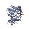
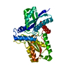
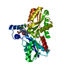
- PDB-2h1v: Crystal structure of the Lys87Ala mutant variant of Bacillus subt... -
+ Open data

- Basic information
| Entry | Database: PDB / ID: 2h1v | ||||||
|---|---|---|---|---|---|---|---|
| Title | Crystal structure of the Lys87Ala mutant variant of Bacillus subtilis ferrochelatase | ||||||
 Components Components | Ferrochelatase | ||||||
 Keywords Keywords | LYASE / ROSSMANN FOLD / PI-HELIX | ||||||
| Function / homology |  Function and homology information Function and homology informationcoproporphyrin ferrochelatase / protoporphyrin ferrochelatase activity / heme biosynthetic process / metal ion binding / cytoplasm Similarity search - Function | ||||||
| Biological species |  | ||||||
| Method |  X-RAY DIFFRACTION / SYNCHROTRON / MOLECULAR REPLACEMENT / Resolution: 1.2 Å X-RAY DIFFRACTION / SYNCHROTRON / MOLECULAR REPLACEMENT / Resolution: 1.2 Å | ||||||
 Authors Authors | Hansson, M.D. / Karlberg, T. / Arys Rahardja, M. / Al-Karadaghi, S. / Hansson, M. | ||||||
 Citation Citation | Journal: Biochemistry / Year: 2007 Title: Amino Acid Residues His183 and Glu264 in Bacillus subtilis Ferrochelatase Direct and Facilitate the Insertion of Metal Ion into Protoporphyrin IX Authors: Hansson, M.D. / Karlberg, T. / Rahardja, M.A. / Al-Karadaghi, S. / Hansson, M. | ||||||
| History |
|
- Structure visualization
| Structure viewer | Molecule: MolmilJmol/JSmol |
|---|
- Downloads & links
-Download
| PDBx/mmCIF format | 2h1v.cif.gz | 162.4 KB | Display | PDBx/mmCIF format |
|---|---|---|---|---|
| PDB format | pdb2h1v.ent.gz | 126.4 KB | Display | PDB format |
| PDBx/mmJSON format | 2h1v.json.gz | Tree view | PDBx/mmJSON format | |
| Others |  Other downloads Other downloads |
-Validation report
| Arichive directory | https://data.pdbj.org/pub/pdb/validation_reports/h1/2h1vftp://data.pdbj.org/pub/pdb/validation_reports/h1/2h1v | HTTPS FTP |
|---|
-Related structure data
| Related structure data |  2h1wC  2hk6C  1dozS S: Starting model for refinement C: citing same article ( |
|---|---|
| Similar structure data |


-Links
 PDBj
PDBj- Assembly
| Deposited unit | 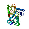
| ||||||||
|---|---|---|---|---|---|---|---|---|---|
| 1 |
| ||||||||
| Unit cell |
|
-Components
| #1: Protein | Mass: 35331.602 Da / Num. of mol.: 1 / Mutation: K87A Source method: isolated from a genetically manipulated source Source: (gene. exp.) | ||
|---|---|---|---|
| #2: Chemical | ChemComp-MG /   Mass: 24.305 Da / Num. of mol.: 4 / Source method: obtained synthetically / Formula: Mg Mass: 24.305 Da / Num. of mol.: 4 / Source method: obtained synthetically / Formula: Mg#3: Water | ChemComp-HOH / |  Mass: 18.015 Da / Num. of mol.: 490 / Source method: isolated from a natural source / Formula: H2O Mass: 18.015 Da / Num. of mol.: 490 / Source method: isolated from a natural source / Formula: H2O |
-Experimental details
-Experiment
| Experiment | Method: X-RAY DIFFRACTION / Number of used crystals: 1 |
|---|
- Sample preparation
| Crystal | Density Matthews: 1.95 Å3/Da / Density % sol: 37.02 % |
|---|---|
| Crystal grow | Temperature: 288 K / Method: vapor diffusion / pH: 7.4 Details: 25-30% PEG 2000, 0.2 M magnesium chloride, 0.1 M Tris-HCl, pH 7.4, VAPOR DIFFUSION, temperature 288K |
-Data collection
| Diffraction | Mean temperature: 100 K |
|---|---|
| Diffraction source | Source: SYNCHROTRON / Site: MAX II  / Beamline: I911-2 / Wavelength: 1.043 / Beamline: I911-2 / Wavelength: 1.043 |
| Detector | Type: MARRESEARCH / Detector: CCD / Date: Nov 18, 2004 |
| Radiation | Monochromator: Bent germanium crystal / Protocol: SINGLE WAVELENGTH / Monochromatic (M) / Laue (L): M / Scattering type: x-ray |
| Radiation wavelength | Wavelength: 1.043 Å / Relative weight: 1 |
| Reflection | Resolution: 1.2→25 Å / Num. all: 83335 / Num. obs: 83273 / % possible obs: 93.9 % / Observed criterion σ(F): 2 / Observed criterion σ(I): 2 / Redundancy: 5.1 % / Rmerge(I) obs: 0.067 / Net I/σ(I): 14 |
| Reflection shell | Resolution: 1.2→1.27 Å / Redundancy: 2.3 % / Rmerge(I) obs: 0.233 / Mean I/σ(I) obs: 5 / Num. unique all: 9813 / % possible all: 70.2 |
- Processing
| Software |
| |||||||||||||||||||||||||||||||||
|---|---|---|---|---|---|---|---|---|---|---|---|---|---|---|---|---|---|---|---|---|---|---|---|---|---|---|---|---|---|---|---|---|---|---|
| Refinement | Method to determine structure: MOLECULAR REPLACEMENT Starting model: 1DOZ Resolution: 1.2→25 Å / Num. parameters: 27732 / Num. restraintsaints: 34600 / Isotropic thermal model: anisotropic / Cross valid method: FREE R / σ(F): 0 / Stereochemistry target values: ENGH AND HUBER Details: ANISOTROPIC REFINEMENT REDUCED FREE R (NO CUTOFF) BY 0.034
| |||||||||||||||||||||||||||||||||
| Refine analyze | Num. disordered residues: 12 / Occupancy sum hydrogen: 0 / Occupancy sum non hydrogen: 2971.5 | |||||||||||||||||||||||||||||||||
| Refinement step | Cycle: LAST / Resolution: 1.2→25 Å
| |||||||||||||||||||||||||||||||||
| Refine LS restraints |
|
