 Movie
Movie Controller

[English] 日本語
 Yorodumi
- PDB-2xtz: Crystal structure of the G alpha protein AtGPA1 from Arabidopsis ... -
+ Open data

- Basic information
| Entry | Database: PDB / ID: 2xtz | ||||||
|---|---|---|---|---|---|---|---|
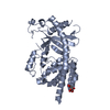
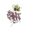
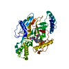
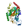
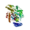
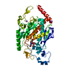
| Title | Crystal structure of the G alpha protein AtGPA1 from Arabidopsis thaliana | ||||||
 Components Components | GUANINE NUCLEOTIDE-BINDING PROTEIN ALPHA-1 SUBUNIT | ||||||
 Keywords Keywords | HYDROLASE / G-PROTEIN SIGNALING / SELF-ACTIVATION / RAS-LIKE DOMAIN | ||||||
| Function / homology |  Function and homology information Function and homology informationprogrammed cell death in response to reactive oxygen species / thylakoid membrane organization / response to low fluence blue light stimulus by blue low-fluence system / positive regulation of abscisic acid-activated signaling pathway / response to pheromone / gibberellic acid mediated signaling pathway / regulation of stomatal closure / L-tyrosine biosynthetic process / blue light signaling pathway / seed germination ...programmed cell death in response to reactive oxygen species / thylakoid membrane organization / response to low fluence blue light stimulus by blue low-fluence system / positive regulation of abscisic acid-activated signaling pathway / response to pheromone / gibberellic acid mediated signaling pathway / regulation of stomatal closure / L-tyrosine biosynthetic process / blue light signaling pathway / seed germination / regulation of stomatal movement / GTPase inhibitor activity / L-phenylalanine biosynthetic process / plasmodesma / abscisic acid-activated signaling pathway / channel regulator activity / sphingosine-1-phosphate receptor signaling pathway / response to glucose / reactive oxygen species metabolic process / G protein-coupled receptor binding / G-protein beta/gamma-subunit complex binding / adenylate cyclase-modulating G protein-coupled receptor signaling pathway / regulation of cell population proliferation / peroxisome / heterotrimeric G-protein complex / GTPase binding / G protein-coupled receptor signaling pathway / GTPase activity / endoplasmic reticulum membrane / GTP binding / metal ion binding / plasma membrane / cytoplasm Similarity search - Function | ||||||
| Biological species |  | ||||||
| Method |  X-RAY DIFFRACTION / SYNCHROTRON / MOLECULAR REPLACEMENT / Resolution: 2.34 Å X-RAY DIFFRACTION / SYNCHROTRON / MOLECULAR REPLACEMENT / Resolution: 2.34 Å | ||||||
 Authors Authors | Jones, J.C. / Duffy, J.W. / Machius, M. / Temple, B.R.S. / Dohlman, H.G. / Jones, A.M. | ||||||
 Citation Citation | Journal: Sci.Signal. / Year: 2011 Title: The Crystal Structure of a Self-Activating G Protein Alpha Subunit Reveals its Distinct Mechanism of Signal Initiation Authors: Jones, J.C. / Duffy, J.W. / Machius, M. / Temple, B.R.S. / Dohlman, H.G. / Jones, A.M. #1: Journal: Proc.Natl.Acad.Sci.USA / Year: 2007 Title: Gtpase Acceleration as the Rate-Limiting Step in Arabidopsis G Protein-Coupled Sugar Signaling. Authors: Johnston, C.A. / Taylor, J.P. / Gao, Y. / Kimple, A.J. / Grigston, J.C. / Chen, J. / Siderovski, D.P. / Jones, A.M. / Willard, F.S. | ||||||
| History |
|
- Structure visualization
| Structure viewer | Molecule: MolmilJmol/JSmol |
|---|
- Downloads & links
-Download
| PDBx/mmCIF format | 2xtz.cif.gz | 409.9 KB | Display | PDBx/mmCIF format |
|---|---|---|---|---|
| PDB format | pdb2xtz.ent.gz | 336.4 KB | Display | PDB format |
| PDBx/mmJSON format | 2xtz.json.gz | Tree view | PDBx/mmJSON format | |
| Others |  Other downloads Other downloads |
-Validation report
| Arichive directory | https://data.pdbj.org/pub/pdb/validation_reports/xt/2xtzftp://data.pdbj.org/pub/pdb/validation_reports/xt/2xtz | HTTPS FTP |
|---|
-Related structure data
| Related structure data | 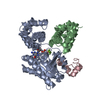 1fqjS S: Starting model for refinement |
|---|---|
| Similar structure data |


-Links
 PDBj
PDBj


- Assembly
| Deposited unit | 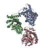
| ||||||||
|---|---|---|---|---|---|---|---|---|---|
| 1 | 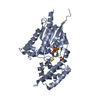
| ||||||||
| 2 | 
| ||||||||
| 3 | 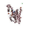
| ||||||||
| Unit cell |
|
-Components
-Protein , 1 types, 3 molecules ABC
| #1: Protein | Mass: 41075.090 Da / Num. of mol.: 3 / Fragment: RESIDUES 37-383 Source method: isolated from a genetically manipulated source Source: (gene. exp.)  |
|---|
-Non-polymers , 5 types, 264 molecules 


| #2: Chemical |  Mass: 24.305 Da / Num. of mol.: 3 / Source method: obtained synthetically / Formula: Mg Mass: 24.305 Da / Num. of mol.: 3 / Source method: obtained synthetically / Formula: Mg#3: Chemical |  Mass: 539.246 Da / Num. of mol.: 3 / Source method: obtained synthetically / Formula: C10H16N5O13P3S Mass: 539.246 Da / Num. of mol.: 3 / Source method: obtained synthetically / Formula: C10H16N5O13P3S#4: Chemical | ChemComp-CL /  Mass: 35.453 Da / Num. of mol.: 4 / Source method: obtained synthetically / Formula: Cl Mass: 35.453 Da / Num. of mol.: 4 / Source method: obtained synthetically / Formula: Cl#5: Chemical | ChemComp-SO4 /  Mass: 96.063 Da / Num. of mol.: 5 / Source method: obtained synthetically / Formula: SO4 Mass: 96.063 Da / Num. of mol.: 5 / Source method: obtained synthetically / Formula: SO4#6: Water | ChemComp-HOH / | Mass: 18.015 Da / Num. of mol.: 249 / Source method: isolated from a natural source / Formula: H2O |
|---|
-Details
| Has protein modification | Y |
|---|---|
| Sequence details | THE 36 N-TERMINAL RESIDUES OF THE FULL-LENGTH PROTEIN WERE REMOVED FOR CRYSTALLIZATION PURPOSES. ...THE 36 N-TERMINAL RESIDUES OF THE FULL-LENGTH PROTEIN WERE REMOVED FOR CRYSTALLIZ |
-Experimental details
-Experiment
| Experiment | Method: X-RAY DIFFRACTION / Number of used crystals: 1 |
|---|
- Sample preparation
| Crystal | Density Matthews: 2.6 Å3/Da / Density % sol: 53.3 % / Description: NONE |
|---|---|
| Crystal grow | Temperature: 277 K / Method: vapor diffusion, hanging drop / pH: 6 Details: CRYSTALS OF ATGPA1 WERE GROWN AT 4 DEGREES CELSIUS USING THE HANGING-DROP VAPOR-DIFFUSION METHOD. DROPS CONTAINED 1.5 UL OF PROTEIN SOLUTION (20 MG/ML ATGPA1 IN 25 MM TRIS-HCL, PH 7.4, 5% ...Details: CRYSTALS OF ATGPA1 WERE GROWN AT 4 DEGREES CELSIUS USING THE HANGING-DROP VAPOR-DIFFUSION METHOD. DROPS CONTAINED 1.5 UL OF PROTEIN SOLUTION (20 MG/ML ATGPA1 IN 25 MM TRIS-HCL, PH 7.4, 5% (V/V) GLYCEROL, 150 MM SODIUM CHLORIDE, 1 MM DTT, 500 UL GTP-GAMMA-S) AND WERE EQUILIBRATED AGAINST 1.5 UL OF 0.3 M MAGNESIUM SULFATE, 0.1 M SODIUM CACODYLATE, PH 6.0, 21% (W/V) PEG 8000. INITIALLY OBTAINED CRYSTALS WERE USED FOR MACROSEEDING. CRYSTALS REACHED THEIR FINAL ROD-SHAPE FORM WITHIN 14 DAYS AFTER MACROSEEDING AND WERE CRYOPROTECTED IN THE MOTHER LIQUOR WITH 8% (V/V) GLYCEROL. |
-Data collection
| Diffraction | Mean temperature: 100 K |
|---|---|
| Diffraction source | Source: SYNCHROTRON / Site: APS  / Beamline: 22-ID / Wavelength: 1 / Beamline: 22-ID / Wavelength: 1 |
| Detector | Type: MARRESEARCH MX300 / Detector: CCD / Date: Nov 16, 2009 / Details: CUSTOM |
| Radiation | Monochromator: SI(111) / Protocol: SINGLE WAVELENGTH / Monochromatic (M) / Laue (L): M / Scattering type: x-ray |
| Radiation wavelength | Wavelength: 1 Å / Relative weight: 1 |
| Reflection | Resolution: 2.34→44.6 Å / Num. obs: 55808 / % possible obs: 100 % / Observed criterion σ(I): -3 / Redundancy: 6 % / Biso Wilson estimate: 52.9 Å2 / Rmerge(I) obs: 0.11 / Net I/σ(I): 17.5 |
| Reflection shell | Resolution: 2.34→2.36 Å / Redundancy: 5.8 % / Rmerge(I) obs: 0.84 / Mean I/σ(I) obs: 1.9 / % possible all: 100 |
- Processing
| Software |
| ||||||||||||||||||||||||||||||||||||||||||||||||||||||||||||||||||||||||||||||||||||||||||||||||||||||||||||||||||||||||||||||||||||||||||||||||||||||||||||||||||||||||||||||||||||||
|---|---|---|---|---|---|---|---|---|---|---|---|---|---|---|---|---|---|---|---|---|---|---|---|---|---|---|---|---|---|---|---|---|---|---|---|---|---|---|---|---|---|---|---|---|---|---|---|---|---|---|---|---|---|---|---|---|---|---|---|---|---|---|---|---|---|---|---|---|---|---|---|---|---|---|---|---|---|---|---|---|---|---|---|---|---|---|---|---|---|---|---|---|---|---|---|---|---|---|---|---|---|---|---|---|---|---|---|---|---|---|---|---|---|---|---|---|---|---|---|---|---|---|---|---|---|---|---|---|---|---|---|---|---|---|---|---|---|---|---|---|---|---|---|---|---|---|---|---|---|---|---|---|---|---|---|---|---|---|---|---|---|---|---|---|---|---|---|---|---|---|---|---|---|---|---|---|---|---|---|---|---|---|---|
| Refinement | Method to determine structure: MOLECULAR REPLACEMENT Starting model: PDB ENTRY 1FQJ Resolution: 2.34→96.03 Å / Cor.coef. Fo:Fc: 0.931 / Cor.coef. Fo:Fc free: 0.904 / SU B: 14.851 / SU ML: 0.164 / Cross valid method: THROUGHOUT / ESU R: 0.295 / ESU R Free: 0.229 / Stereochemistry target values: MAXIMUM LIKELIHOOD Details: HYDROGENS HAVE BEEN ADDED IN THE RIDING POSITIONS. ATOM RECORD CONTAINS SUM OF TLS AND RESIDUAL B FACTORS
| ||||||||||||||||||||||||||||||||||||||||||||||||||||||||||||||||||||||||||||||||||||||||||||||||||||||||||||||||||||||||||||||||||||||||||||||||||||||||||||||||||||||||||||||||||||||
| Solvent computation | Ion probe radii: 0.8 Å / Shrinkage radii: 0.8 Å / VDW probe radii: 1.4 Å / Solvent model: MASK | ||||||||||||||||||||||||||||||||||||||||||||||||||||||||||||||||||||||||||||||||||||||||||||||||||||||||||||||||||||||||||||||||||||||||||||||||||||||||||||||||||||||||||||||||||||||
| Displacement parameters | Biso mean: 44.512 Å2
| ||||||||||||||||||||||||||||||||||||||||||||||||||||||||||||||||||||||||||||||||||||||||||||||||||||||||||||||||||||||||||||||||||||||||||||||||||||||||||||||||||||||||||||||||||||||
| Refinement step | Cycle: LAST / Resolution: 2.34→96.03 Å
| ||||||||||||||||||||||||||||||||||||||||||||||||||||||||||||||||||||||||||||||||||||||||||||||||||||||||||||||||||||||||||||||||||||||||||||||||||||||||||||||||||||||||||||||||||||||
| Refine LS restraints |
|