 Movie
Movie Controller

[English] 日本語
 Yorodumi
- PDB-1ztb: Crystal Structure of Chorismate Synthase from Mycobacterium tuber... -
+ Open data

- Basic information
| Entry | Database: PDB / ID: 1ztb | ||||||
|---|---|---|---|---|---|---|---|
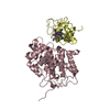
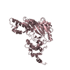
| Title | Crystal Structure of Chorismate Synthase from Mycobacterium tuberculosis | ||||||
 Components Components | Chorismate synthase | ||||||
 Keywords Keywords | LIGASE / BETA-ALPHA-BETA / FLAVOPROTEIN / SHIKIMATE | ||||||
| Function / homology |  Function and homology information Function and homology informationchorismate synthase / chorismate synthase activity / Chorismate via Shikimate Pathway / chorismate biosynthetic process / aromatic amino acid biosynthetic process / amino acid biosynthetic process / oxidoreductase activity, acting on NAD(P)H / NAD binding / FMN binding / cytosol Similarity search - Function | ||||||
| Biological species |   Mycobacterium tuberculosis (bacteria) Mycobacterium tuberculosis (bacteria) | ||||||
| Method |  X-RAY DIFFRACTION / SYNCHROTRON / MOLECULAR REPLACEMENT / Resolution: 2.65 Å X-RAY DIFFRACTION / SYNCHROTRON / MOLECULAR REPLACEMENT / Resolution: 2.65 Å | ||||||
 Authors Authors | Dias, M.V.B. / Borges, J.C. / Ely, F. / Pereira, J.H. / Canduri, F. / Ramos, C.H.I. / Frazzon, J. / Palma, M.S. / Basso, L.A. / Santos, D.S. / Azevedo Jr., W.F. | ||||||
 Citation Citation | Journal: J.Struct.Biol. / Year: 2006 Title: Structure of chorismate synthase from Mycobacterium tuberculosis Authors: Dias, M.V.B. / Borges, J.C. / Ely, F. / Pereira, J.H. / Canduri, F. / Ramos, C.H.I. / Frazzon, J. / Palma, M.S. / Basso, L.A. / Santos, D.S. / de Azevedo Jr., W.F. | ||||||
| History |
|
- Structure visualization
| Structure viewer | Molecule: MolmilJmol/JSmol |
|---|
- Downloads & links
-Download
| PDBx/mmCIF format | 1ztb.cif.gz | 89.6 KB | Display | PDBx/mmCIF format |
|---|---|---|---|---|
| PDB format | pdb1ztb.ent.gz | 68 KB | Display | PDB format |
| PDBx/mmJSON format | 1ztb.json.gz | Tree view | PDBx/mmJSON format | |
| Others |  Other downloads Other downloads |
-Validation report
| Arichive directory | https://data.pdbj.org/pub/pdb/validation_reports/zt/1ztbftp://data.pdbj.org/pub/pdb/validation_reports/zt/1ztb | HTTPS FTP |
|---|
-Related structure data
| Related structure data |  1qxoS S: Starting model for refinement |
|---|---|
| Similar structure data |


-Links
 PDBj
PDBj
- Assembly
| Deposited unit | 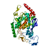
| ||||||||||
|---|---|---|---|---|---|---|---|---|---|---|---|
| 1 |
| ||||||||||
| 2 | 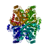
| ||||||||||
| Unit cell |
| ||||||||||
| Components on special symmetry positions |
|
-Components
| #1: Protein | Mass: 41844.383 Da / Num. of mol.: 1 Source method: isolated from a genetically manipulated source Source: (gene. exp.) Mycobacterium tuberculosis (bacteria) / Gene: aroC, aroF / Plasmid: pET-23a(+) / Production host: References: UniProt: P63611, UniProt: P9WPY1*PLUS, chorismate synthase |
|---|---|
| #2: Water | ChemComp-HOH /  Mass: 18.015 Da / Num. of mol.: 243 / Source method: isolated from a natural source / Formula: H2O Mass: 18.015 Da / Num. of mol.: 243 / Source method: isolated from a natural source / Formula: H2O |
-Experimental details
-Experiment
| Experiment | Method: X-RAY DIFFRACTION / Number of used crystals: 1 |
|---|
- Sample preparation
| Crystal | Density Matthews: 4.55 Å3/Da / Density % sol: 72.95 % |
|---|---|
| Crystal grow | Temperature: 298 K / Method: vapor diffusion, hanging drop / pH: 7.8 Details: PEG 400, magnesium chloride, Hepes, pH 7.8, VAPOR DIFFUSION, HANGING DROP, temperature 298K |
-Data collection
| Diffraction | Mean temperature: 100 K |
|---|---|
| Diffraction source | Source: SYNCHROTRON / Site: LNLS  / Beamline: D03B-MX1 / Wavelength: 1.427 Å / Beamline: D03B-MX1 / Wavelength: 1.427 Å |
| Detector | Type: MARRESEARCH / Detector: CCD / Date: Mar 22, 2004 |
| Radiation | Monochromator: GRAPHITE / Protocol: SINGLE WAVELENGTH / Monochromatic (M) / Laue (L): M / Scattering type: x-ray |
| Radiation wavelength | Wavelength: 1.427 Å / Relative weight: 1 |
| Reflection | Resolution: 2.65→52.93 Å / Num. all: 22658 / Num. obs: 22383 / % possible obs: 97.3 % / Observed criterion σ(F): 2 / Observed criterion σ(I): 2 / Rmerge(I) obs: 0.07 |
| Reflection shell | Resolution: 2.65→2.79 Å / Rmerge(I) obs: 0.337 / Num. unique all: 3129 / % possible all: 95.2 |
- Processing
| Software |
| ||||||||||||||||||||||||||||||||||||||||||||||||||||||||||||||||||||||||||||||||||||||||||
|---|---|---|---|---|---|---|---|---|---|---|---|---|---|---|---|---|---|---|---|---|---|---|---|---|---|---|---|---|---|---|---|---|---|---|---|---|---|---|---|---|---|---|---|---|---|---|---|---|---|---|---|---|---|---|---|---|---|---|---|---|---|---|---|---|---|---|---|---|---|---|---|---|---|---|---|---|---|---|---|---|---|---|---|---|---|---|---|---|---|---|---|
| Refinement | Method to determine structure: MOLECULAR REPLACEMENT Starting model: PDB ENTRY 1QXO Resolution: 2.65→52.93 Å / Cor.coef. Fo:Fc: 0.956 / Cor.coef. Fo:Fc free: 0.925 / SU B: 7.277 / SU ML: 0.148 / Cross valid method: THROUGHOUT / σ(F): 2 / ESU R: 0.284 / ESU R Free: 0.241 / Stereochemistry target values: MAXIMUM LIKELIHOOD / Details: HYDROGENS HAVE BEEN ADDED IN THE RIDING POSITIONS
| ||||||||||||||||||||||||||||||||||||||||||||||||||||||||||||||||||||||||||||||||||||||||||
| Solvent computation | Ion probe radii: 0.8 Å / Shrinkage radii: 0.8 Å / VDW probe radii: 1.2 Å / Solvent model: MASK | ||||||||||||||||||||||||||||||||||||||||||||||||||||||||||||||||||||||||||||||||||||||||||
| Displacement parameters | Biso mean: 34.902 Å2
| ||||||||||||||||||||||||||||||||||||||||||||||||||||||||||||||||||||||||||||||||||||||||||
| Refinement step | Cycle: LAST / Resolution: 2.65→52.93 Å
| ||||||||||||||||||||||||||||||||||||||||||||||||||||||||||||||||||||||||||||||||||||||||||
| Refine LS restraints |
| ||||||||||||||||||||||||||||||||||||||||||||||||||||||||||||||||||||||||||||||||||||||||||
| LS refinement shell | Resolution: 2.65→2.719 Å / Total num. of bins used: 20
|
