 Movie
Movie Controller

[English] 日本語
 Yorodumi
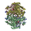
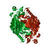
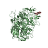
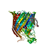
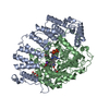
- PDB-2o12: Mycobacterium tuberculosis Chorismate synthase in complex with FMN -
+ Open data

- Basic information
| Entry | Database: PDB / ID: 2o12 | ||||||
|---|---|---|---|---|---|---|---|
| Title | Mycobacterium tuberculosis Chorismate synthase in complex with FMN | ||||||
 Components Components | Chorismate synthase | ||||||
 Keywords Keywords | LYASE / Shikimate pathway / M. tuberculosis / Chorismate synthase | ||||||
| Function / homology |  Function and homology information Function and homology informationchorismate synthase / chorismate synthase activity / Chorismate via Shikimate Pathway / chorismate biosynthetic process / aromatic amino acid biosynthetic process / amino acid biosynthetic process / oxidoreductase activity, acting on NAD(P)H / NAD binding / FMN binding / cytosol Similarity search - Function | ||||||
| Biological species |   Mycobacterium tuberculosis (bacteria) Mycobacterium tuberculosis (bacteria) | ||||||
| Method |  X-RAY DIFFRACTION / SYNCHROTRON / FOURIER SYNTHESIS / Resolution: 1.72 Å X-RAY DIFFRACTION / SYNCHROTRON / FOURIER SYNTHESIS / Resolution: 1.72 Å | ||||||
 Authors Authors | Bruning, M. / Bartunik, H.D. | ||||||
 Citation Citation | Journal: To be Published Title: Complexes of Chorismate synthase from M. tuberculosis Authors: Bruning, M. / Bourenkov, G.P. / Strizhov, N.I. / Bartunik, H.D. | ||||||
| History |
|
- Structure visualization
| Structure viewer | Molecule: MolmilJmol/JSmol |
|---|
- Downloads & links
-Download
| PDBx/mmCIF format | 2o12.cif.gz | 102.8 KB | Display | PDBx/mmCIF format |
|---|---|---|---|---|
| PDB format | pdb2o12.ent.gz | 77.1 KB | Display | PDB format |
| PDBx/mmJSON format | 2o12.json.gz | Tree view | PDBx/mmJSON format | |
| Others |  Other downloads Other downloads |
-Validation report
| Arichive directory | https://data.pdbj.org/pub/pdb/validation_reports/o1/2o12ftp://data.pdbj.org/pub/pdb/validation_reports/o1/2o12 | HTTPS FTP |
|---|
-Related structure data


-Links
 PDBj
PDBj
- Assembly
| Deposited unit | 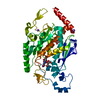
| |||||||||
|---|---|---|---|---|---|---|---|---|---|---|
| 1 | 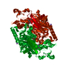
| |||||||||
| 2 | 
| |||||||||
| Unit cell |
| |||||||||
| Components on special symmetry positions |
|
-Components
| #1: Protein | Mass: 42673.250 Da / Num. of mol.: 1 Source method: isolated from a genetically manipulated source Source: (gene. exp.) Mycobacterium tuberculosis (bacteria) / Strain: H37Rv / Gene: aroF / Production host: References: UniProt: P63611, UniProt: P9WPY1*PLUS, chorismate synthase | ||||||
|---|---|---|---|---|---|---|---|
| #2: Chemical | ChemComp-ACT /   Mass: 59.044 Da / Num. of mol.: 4 / Source method: obtained synthetically / Formula: C2H3O2 Mass: 59.044 Da / Num. of mol.: 4 / Source method: obtained synthetically / Formula: C2H3O2#3: Chemical |   Mass: 22.990 Da / Num. of mol.: 2 / Source method: obtained synthetically / Formula: Na Mass: 22.990 Da / Num. of mol.: 2 / Source method: obtained synthetically / Formula: Na#4: Chemical | ChemComp-FMN / |   Mass: 456.344 Da / Num. of mol.: 1 / Source method: obtained synthetically / Formula: C17H21N4O9P Mass: 456.344 Da / Num. of mol.: 1 / Source method: obtained synthetically / Formula: C17H21N4O9P#5: Water | ChemComp-HOH / |  Mass: 18.015 Da / Num. of mol.: 511 / Source method: isolated from a natural source / Formula: H2O Mass: 18.015 Da / Num. of mol.: 511 / Source method: isolated from a natural source / Formula: H2O |
-Experimental details
-Experiment
| Experiment | Method: X-RAY DIFFRACTION / Number of used crystals: 1 |
|---|
- Sample preparation
| Crystal | Density Matthews: 4.77 Å3/Da / Density % sol: 74.24 % |
|---|---|
| Crystal grow | Temperature: 291 K / Method: vapor diffusion, sitting drop / pH: 4.5 Details: 4.8 M NH4-Acetate, 0.1 M Na-Acetate, pH 4.5, VAPOR DIFFUSION, SITTING DROP, temperature 291K |
-Data collection
| Diffraction | Mean temperature: 100 K |
|---|---|
| Diffraction source | Source: SYNCHROTRON / Site: MPG/DESY, HAMBURG  / Beamline: BW6 / Wavelength: 1.05 Å / Beamline: BW6 / Wavelength: 1.05 Å |
| Detector | Type: MAR CCD 165 mm / Detector: CCD / Date: Nov 3, 2005 / Details: Au-coated planar; toroidal mirrors |
| Radiation | Monochromator: Si111 / Protocol: SINGLE WAVELENGTH / Monochromatic (M) / Laue (L): M / Scattering type: x-ray |
| Radiation wavelength | Wavelength: 1.05 Å / Relative weight: 1 |
| Reflection | Resolution: 1.72→20 Å / Num. all: 88402 / Num. obs: 88273 / % possible obs: 99.9 % / Observed criterion σ(I): 2.3 / Redundancy: 7 % / Biso Wilson estimate: 25.9 Å2 / Rmerge(I) obs: 0.086 / Net I/σ(I): 20.6 |
| Reflection shell | Resolution: 1.72→1.74 Å / Redundancy: 7 % / Rmerge(I) obs: 0.932 / Mean I/σ(I) obs: 2.4 / Num. unique all: 3488 / % possible all: 99.9 |
- Processing
| Software |
| ||||||||||||||||||||||||||||||||||||||||||||||||||||||||||||||||||||||||||||||||||||||||||||||||||||||||||||||||||||||||||||||||||
|---|---|---|---|---|---|---|---|---|---|---|---|---|---|---|---|---|---|---|---|---|---|---|---|---|---|---|---|---|---|---|---|---|---|---|---|---|---|---|---|---|---|---|---|---|---|---|---|---|---|---|---|---|---|---|---|---|---|---|---|---|---|---|---|---|---|---|---|---|---|---|---|---|---|---|---|---|---|---|---|---|---|---|---|---|---|---|---|---|---|---|---|---|---|---|---|---|---|---|---|---|---|---|---|---|---|---|---|---|---|---|---|---|---|---|---|---|---|---|---|---|---|---|---|---|---|---|---|---|---|---|---|
| Refinement | Method to determine structure: FOURIER SYNTHESIS / Resolution: 1.72→19.74 Å / Cor.coef. Fo:Fc: 0.971 / Cor.coef. Fo:Fc free: 0.964 / SU B: 1.401 / SU ML: 0.046 / Cross valid method: THROUGHOUT / ESU R: 0.065 / ESU R Free: 0.068 / Stereochemistry target values: MAXIMUM LIKELIHOOD / Details: HYDROGENS HAVE BEEN ADDED IN THE RIDING POSITIONS
| ||||||||||||||||||||||||||||||||||||||||||||||||||||||||||||||||||||||||||||||||||||||||||||||||||||||||||||||||||||||||||||||||||
| Solvent computation | Ion probe radii: 0.8 Å / Shrinkage radii: 0.8 Å / VDW probe radii: 1.2 Å / Solvent model: BABINET MODEL WITH MASK | ||||||||||||||||||||||||||||||||||||||||||||||||||||||||||||||||||||||||||||||||||||||||||||||||||||||||||||||||||||||||||||||||||
| Displacement parameters | Biso mean: 23.079 Å2
| ||||||||||||||||||||||||||||||||||||||||||||||||||||||||||||||||||||||||||||||||||||||||||||||||||||||||||||||||||||||||||||||||||
| Refinement step | Cycle: LAST / Resolution: 1.72→19.74 Å
| ||||||||||||||||||||||||||||||||||||||||||||||||||||||||||||||||||||||||||||||||||||||||||||||||||||||||||||||||||||||||||||||||||
| Refine LS restraints |
| ||||||||||||||||||||||||||||||||||||||||||||||||||||||||||||||||||||||||||||||||||||||||||||||||||||||||||||||||||||||||||||||||||
| LS refinement shell | Resolution: 1.72→1.764 Å / Total num. of bins used: 20
|
