tRNA wobble uridine modification / tRNA methylation / Hydrolases; Acting on acid anhydrides; Acting on GTP to facilitate cellular and subcellular movement / GTPase activity / GTP binding / metal ion binding / identical protein binding / cytosol / cytoplasm Similarity search - Function
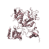
The biological assembly is a homodimer (obtained by superimposition of the full-length molecule (A) and the N-terminal domain (B)). Dimerisation via the N-terminal domain is essential for the formation of the THF-binding site.
-
Components
#1: Protein
ProbabletRNAmodificationGTPasetrmE / TrmE GTP-binding protein
Mass: 54189.203 Da / Num. of mol.: 1 Source method: isolated from a genetically manipulated source Source: (gene. exp.) Thermotoga maritima (bacteria) / Gene: TrmE / Plasmid: pET20b / Production host: Escherichia coli (E. coli) / Strain (production host): BL21DE3 (TrmE-) / References: UniProt: Q9WYA4
#2: Protein
ProbabletRNAmodificationGTPasetrmE / TrmE GTP-binding protein
Mass: 16495.090 Da / Num. of mol.: 1 / Fragment: N-terminal domain (residues 1-118) Source method: isolated from a genetically manipulated source Source: (gene. exp.) Thermotoga maritima (bacteria) / Gene: TrmE / Plasmid: pET20b / Production host: Escherichia coli (E. coli) / Strain (production host): BL21DE3 (TrmE-) / References: UniProt: Q9WYA4
Protocol: SINGLE WAVELENGTH / Monochromatic (M) / Laue (L): M / Scattering type: x-ray
Radiation wavelength
Wavelength: 0.934 Å / Relative weight: 1
Reflection
Resolution: 2.3→19.7 Å / Num. all: 44838 / Num. obs: 44730 / % possible obs: 99.8 % / Observed criterion σ(F): 0 / Redundancy: 6 % / Biso Wilson estimate: 37.1 Å2 / Rmerge(I) obs: 0.065 / Net I/σ(I): 15.5
Reflection shell
Resolution: 2.3→2.4 Å / Redundancy: 6 % / Rmerge(I) obs: 0.44 / Mean I/σ(I) obs: 4.05 / Num. unique all: 5335 / % possible all: 99.7
-
Processing
Software
Name
Classification
XDS
datascaling
XSCALE
datascaling
SHARP
phasing
CNS
refinement
XDS
datareduction
Refinement
Method to determine structure: SAD / Resolution: 2.3→19.7 Å / Isotropic thermal model: RESTRAINED / Cross valid method: THROUGHOUT / σ(F): 0 / Stereochemistry target values: Engh & Huber Details: THe authors are not sure about the position of the two SO4 ions with the high B-factors. From the density shape they would expect this type of Ion there due to the Ammonium Sulphate condition.
Rfactor
Num. reflection
% reflection
Selection details
Rfree
0.252
2237
-
RANDOM
Rwork
0.226
-
-
-
all
0.227
44838
-
-
obs
0.227
44730
99.6 %
-
Displacement parameters
Biso mean: 60.4 Å2
Baniso -1
Baniso -2
Baniso -3
1-
-10.55 Å2
-1.34 Å2
0 Å2
2-
-
-10.55 Å2
0 Å2
3-
-
-
21.1 Å2
Refine analyze
Free
Obs
Luzzati coordinate error
0.34 Å
0.3 Å
Luzzati d res low
-
5 Å
Luzzati sigma a
0.32 Å
0.3 Å
Refinement step
Cycle: LAST / Resolution: 2.3→19.7 Å
Protein
Nucleic acid
Ligand
Solvent
Total
Num. atoms
4546
0
25
137
4708
Refine LS restraints
Refine-ID
Type
Dev ideal
X-RAY DIFFRACTION
x_bond_d
0.006
X-RAY DIFFRACTION
x_angle_deg
1.3
X-RAY DIFFRACTION
x_torsion_deg
22.3
X-RAY DIFFRACTION
x_torsion_impr_deg
0.88
+
About Yorodumi
-
News
-
Feb 9, 2022. New format data for meta-information of EMDB entries
New format data for meta-information of EMDB entries
Version 3 of the EMDB header file is now the official format.
The previous official version 1.9 will be removed from the archive.
In the structure databanks used in Yorodumi, some data are registered as the other names, "COVID-19 virus" and "2019-nCoV". Here are the details of the virus and the list of structure data.
Jan 31, 2019. EMDB accession codes are about to change! (news from PDBe EMDB page)
EMDB accession codes are about to change! (news from PDBe EMDB page)
The allocation of 4 digits for EMDB accession codes will soon come to an end. Whilst these codes will remain in use, new EMDB accession codes will include an additional digit and will expand incrementally as the available range of codes is exhausted. The current 4-digit format prefixed with “EMD-” (i.e. EMD-XXXX) will advance to a 5-digit format (i.e. EMD-XXXXX), and so on. It is currently estimated that the 4-digit codes will be depleted around Spring 2019, at which point the 5-digit format will come into force.
The EM Navigator/Yorodumi systems omit the EMD- prefix.
Related info.:Q: What is EMD? / ID/Accession-code notation in Yorodumi/EM Navigator
Yorodumi is a browser for structure data from EMDB, PDB, SASBDB, etc.
This page is also the successor to EM Navigator detail page, and also detail information page/front-end page for Omokage search.
The word "yorodu" (or yorozu) is an old Japanese word meaning "ten thousand". "mi" (miru) is to see.
Related info.:EMDB / PDB / SASBDB / Comparison of 3 databanks / Yorodumi Search / Aug 31, 2016. New EM Navigator & Yorodumi / Yorodumi Papers / Jmol/JSmol / Function and homology information / Changes in new EM Navigator and Yorodumi
 Movie
Movie Controller

 Yorodumi
 Open data
 Basic information
 Components
 Keywords
 Function and homology information
 Thermotoga maritima (bacteria)
 X-RAY DIFFRACTION /
  Authors
 Citation
 Structure visualization
 Downloads & links
 Other downloads


 PDBj
PDBj Assembly


 Mass: 96.063 Da / Num. of mol.: 5 / Source method: obtained synthetically / Formula: SO4
 Mass: 18.015 Da / Num. of mol.: 137 / Source method: isolated from a natural source / Formula: H2O
 Sample preparation
 / Beamline: ID14-1 / Wavelength: 0.934 Å
 Processing