 Movie
Movie Controller


+ Open data

- Basic information
| Entry | Database: EMDB / ID: EMD-8849 | |||||||||
|---|---|---|---|---|---|---|---|---|---|---|
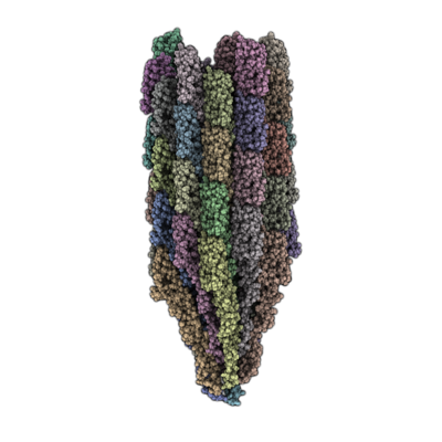
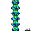
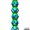
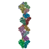
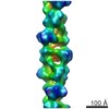
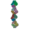
| Title | Cryo-EM structure of B. subtilis flagellar filaments A233V | |||||||||
 Map data Map data | Cryo-EM structure of B. subtilis flagellar filaments A233V | |||||||||
 Sample Sample |
| |||||||||
 Keywords Keywords | bacteria flagella / helical polymers / cryo-EM / PROTEIN FIBRIL | |||||||||
| Function / homology | Flagellin, C-terminal domain / Bacterial flagellin C-terminal helical region / Flagellin / Flagellin, N-terminal domain / Bacterial flagellin N-terminal helical region / bacterial-type flagellum / structural molecule activity / extracellular region / Flagellin Function and homology information Function and homology information | |||||||||
| Biological species |  | |||||||||
| Method | helical reconstruction / cryo EM / negative staining / Resolution: 5.5 Å | |||||||||
 Authors Authors | Wang F / Burrage AM | |||||||||
| Funding support |  United States, 2 items United States, 2 items
| |||||||||
 Citation Citation | Journal: Nat Commun / Year: 2017 Title: A structural model of flagellar filament switching across multiple bacterial species. Authors: Fengbin Wang / Andrew M Burrage / Sandra Postel / Reece E Clark / Albina Orlova / Eric J Sundberg / Daniel B Kearns / Edward H Egelman / Abstract: The bacterial flagellar filament has long been studied to understand how a polymer composed of a single protein can switch between different supercoiled states with high cooperativity. Here we ...The bacterial flagellar filament has long been studied to understand how a polymer composed of a single protein can switch between different supercoiled states with high cooperativity. Here we present near-atomic resolution cryo-EM structures for flagellar filaments from both Gram-positive Bacillus subtilis and Gram-negative Pseudomonas aeruginosa. Seven mutant flagellar filaments in B. subtilis and two in P. aeruginosa capture two different states of the filament. These reliable atomic models of both states reveal conserved molecular interactions in the interior of the filament among B. subtilis, P. aeruginosa and Salmonella enterica. Using the detailed information about the molecular interactions in two filament states, we successfully predict point mutations that shift the equilibrium between those two states. Further, we observe the dimerization of P. aeruginosa outer domains without any perturbation of the conserved interior of the filament. Our results give new insights into how the flagellin sequence has been "tuned" over evolution.Bacterial flagellar filaments are composed almost entirely of a single protein-flagellin-which can switch between different supercoiled states in a highly cooperative manner. Here the authors present near-atomic resolution cryo-EM structures of nine flagellar filaments, and begin to shed light on the molecular basis of filament switching. | |||||||||
| History |
|
- Structure visualization
| Movie |
Movie viewer |
|---|---|
| Structure viewer | EM map: SurfViewMolmilJmol/JSmol |
| Supplemental images |
- Downloads & links
-EMDB archive
| Map data | emd_8849.map.gz | 29.1 MB | EMDB map data format | |
|---|---|---|---|---|
| Header (meta data) | emd-8849-v30.xmlemd-8849.xml | 13.1 KB 13.1 KB | Display Display | EMDB header |
| Images |  emd_8849.png emd_8849.png | 113.2 KB | ||
| Filedesc metadata | emd-8849.cif.gz | 5.5 KB | ||
| Archive directory |  http://ftp.pdbj.org/pub/emdb/structures/EMD-8849ftp://ftp.pdbj.org/pub/emdb/structures/EMD-8849 http://ftp.pdbj.org/pub/emdb/structures/EMD-8849ftp://ftp.pdbj.org/pub/emdb/structures/EMD-8849 | HTTPS FTP |
-Related structure data
| Related structure data | 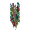 5wjvMC 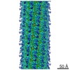 8847C 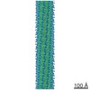 8848C 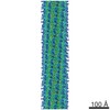 8850C 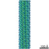 8851C 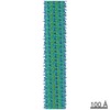 8852C 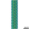 8853C 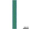 8855C 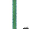 8856C 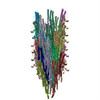 5wjtC 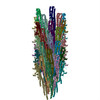 5wjuC 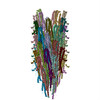 5wjwC 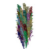 5wjxC 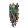 5wjyC 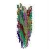 5wjzC 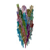 5wk5C 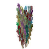 5wk6C C: citing same article ( M: atomic model generated by this map |
|---|---|
| Similar structure data |


-Links
| EMDB pages | EMDB (EBI/PDBe) / EMDataResource |
|---|
-Map
| File | Download / File: emd_8849.map.gz / Format: CCP4 / Size: 512 MB / Type: IMAGE STORED AS FLOATING POINT NUMBER (4 BYTES) | ||||||||||||||||||||||||||||||||||||||||||||||||||||||||||||
|---|---|---|---|---|---|---|---|---|---|---|---|---|---|---|---|---|---|---|---|---|---|---|---|---|---|---|---|---|---|---|---|---|---|---|---|---|---|---|---|---|---|---|---|---|---|---|---|---|---|---|---|---|---|---|---|---|---|---|---|---|---|
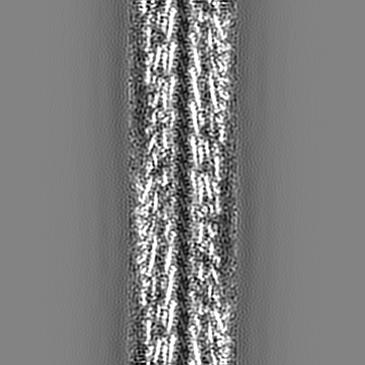
| Annotation | Cryo-EM structure of B. subtilis flagellar filaments A233V | ||||||||||||||||||||||||||||||||||||||||||||||||||||||||||||
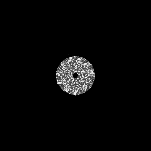
| Projections & slices | Image control
Images are generated by Spider. | ||||||||||||||||||||||||||||||||||||||||||||||||||||||||||||
| Voxel size | X=Y=Z: 1.05 Å | ||||||||||||||||||||||||||||||||||||||||||||||||||||||||||||
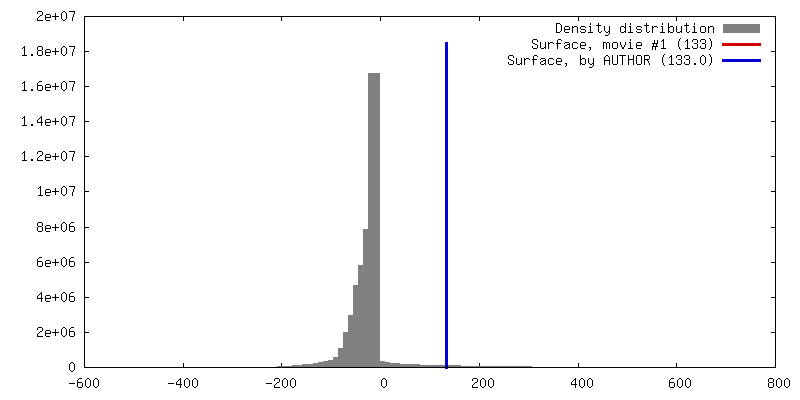
| Density |
| ||||||||||||||||||||||||||||||||||||||||||||||||||||||||||||
| Symmetry | Space group: 1 | ||||||||||||||||||||||||||||||||||||||||||||||||||||||||||||
| Details | EMDB XML:
CCP4 map header:
| ||||||||||||||||||||||||||||||||||||||||||||||||||||||||||||
 Z (Sec.)
 Y (Row.)
 X (Col.)


-Supplemental data
- Sample components
-Entire : Bacillus subtilis flagella filament
| Entire | Name: Bacillus subtilis flagella filament |
|---|---|
| Components |
|
-Supramolecule #1: Bacillus subtilis flagella filament
| Supramolecule | Name: Bacillus subtilis flagella filament / type: complex / ID: 1 / Parent: 0 / Macromolecule list: all |
|---|---|
| Source (natural) | Organism: |
-Macromolecule #1: Flagellin
| Macromolecule | Name: Flagellin / type: protein_or_peptide / ID: 1 / Number of copies: 46 / Enantiomer: LEVO |
|---|---|
| Source (natural) | Organism: |
| Molecular weight | Theoretical: 32.689336 KDa |
| Recombinant expression | Organism: |
| Sequence | String: MRINHNIAAL NTLNRLSSNN SASQKNMEKL SSGLRINRAG DDAAGLAISE KMRGQIRGLE MASKNSQDGI SLIQTAEGAL TETHAILQR VRELVVQAGN TGTQDKATDL QSIQDEISAL TDEIDGISNR TEFNGKKLLD GTYKVDTATP ANQKNLVFQI G ANATQQIS ...String: MRINHNIAAL NTLNRLSSNN SASQKNMEKL SSGLRINRAG DDAAGLAISE KMRGQIRGLE MASKNSQDGI SLIQTAEGAL TETHAILQR VRELVVQAGN TGTQDKATDL QSIQDEISAL TDEIDGISNR TEFNGKKLLD GTYKVDTATP ANQKNLVFQI G ANATQQIS VNIEDMGADA LGIKEADGSI AALHSVNDLD VTKFADNAAD CADIGFDAQL KVVDEAINQV SSQRVKLGAV QN RLEHTIN NLSASGENLT AAESRIRDVD MAKEMSEFTK NNILSQASQA MLAQANQQPQ NVLQLLR UniProtKB: Flagellin |
-Experimental details
-Structure determination
| Method | negative staining, cryo EM |
|---|---|
 Processing Processing | helical reconstruction |
| Aggregation state | filament |
-Sample preparation
| Concentration | 0.1 mg/mL |
|---|---|
| Buffer | pH: 6.8 / Details: Imidazole buffer |
| Staining | Type: NEGATIVE / Material: negative stain |
| Grid | Pretreatment - Type: PLASMA CLEANING |
| Vitrification | Cryogen name: ETHANE / Chamber humidity: 90 % |
- Electron microscopy
| Microscope | FEI TITAN KRIOS |
|---|---|
| Image recording | Film or detector model: FEI FALCON II (4k x 4k) / Detector mode: INTEGRATING / Average exposure time: 3.0 sec. / Average electron dose: 20.0 e/Å2 Details: Images were stored containing seven parts, where each part represented a set of frames corresponding to a dose of ~20 electrons per Angstrom^2. The full dose image stack was used for the ...Details: Images were stored containing seven parts, where each part represented a set of frames corresponding to a dose of ~20 electrons per Angstrom^2. The full dose image stack was used for the estimation of the CTF as well as for boxing filaments. Only the first two parts were used for the reconstruction (~5 electrons per Angstrom^2). |
| Electron beam | Acceleration voltage: 300 kV / Electron source:  FIELD EMISSION GUN FIELD EMISSION GUN |
| Electron optics | Illumination mode: FLOOD BEAM / Imaging mode: BRIGHT FIELD |
| Experimental equipment |  Model: Titan Krios / Image courtesy: FEI Company |
-Image processing
| Final reconstruction | Applied symmetry - Helical parameters - Δz: 4.64 Å Applied symmetry - Helical parameters - Δ&Phi: 65.81 ° Applied symmetry - Helical parameters - Axial symmetry: C1 (asymmetric) Algorithm: BACK PROJECTION / Resolution.type: BY AUTHOR / Resolution: 5.5 Å / Resolution method: OTHER / Software - Name: SPIDER / Details: model-map FSC 0.38 cut-off / Number images used: 33992 |
|---|---|
| Startup model | Type of model: OTHER / Details: featureless cylinder |
| Final angle assignment | Type: NOT APPLICABLE / Software - Name: SPIDER |
-Atomic model buiding 1
| Refinement | Space: REAL |
|---|---|
| Output model | PDB-5wjv: |
