 Movie
Movie Controller

[English] 日本語
 Yorodumi
- EMDB-1313: Post-translational cleavage of recombinantly expressed nitrilase ... -
+ Open data

- Basic information
| Entry | Database: EMDB / ID: EMD-1313 | |||||||||
|---|---|---|---|---|---|---|---|---|---|---|
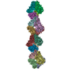
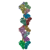
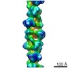
| Title | Post-translational cleavage of recombinantly expressed nitrilase from Rhodococcus rhodochrous J1 yields a stable, active helical form. | |||||||||
 Map data Map data | Map of the carboxy-terminal truncated nitrilase helix from Rhodococcus rhodochrous J1 | |||||||||
 Sample Sample |
| |||||||||
| Function / homology | nitrilase activity / Carbon-nitrogen hydrolase Function and homology information Function and homology information | |||||||||
| Biological species |  Rhodococcus rhodochrous (bacteria) Rhodococcus rhodochrous (bacteria) | |||||||||
| Method | helical reconstruction / negative staining / Resolution: 18.0 Å | |||||||||
 Authors Authors | Thuku RN / Weber BW / Varsani A / Sewell BT | |||||||||
 Citation Citation | Journal: FEBS J / Year: 2007 Title: Post-translational cleavage of recombinantly expressed nitrilase from Rhodococcus rhodochrous J1 yields a stable, active helical form. Authors: R Ndoria Thuku / Brandon W Weber / Arvind Varsani / B Trevor Sewell /  Abstract: Nitrilases convert nitriles to the corresponding carboxylic acids and ammonia. The nitrilase from Rhodococcus rhodochrous J1 is known to be inactive as a dimer but to become active on oligomerization. ...Nitrilases convert nitriles to the corresponding carboxylic acids and ammonia. The nitrilase from Rhodococcus rhodochrous J1 is known to be inactive as a dimer but to become active on oligomerization. The recombinant enzyme undergoes post-translational cleavage at approximately residue 327, resulting in the formation of active, helical homo-oligomers. Determining the 3D structure of these helices using electron microscopy, followed by fitting the stain envelope with a model based on homology with other members of the nitrilase superfamily, enables the interacting surfaces to be identified. This also suggests that the reason for formation of the helices is related to the removal of steric hindrance arising from the 39 C-terminal amino acids from the wild-type protein. The helical form can be generated by expressing only residues 1-327. | |||||||||
| History |
|
- Structure visualization
| Movie |
Movie viewer |
|---|---|
| Structure viewer | EM map: SurfViewMolmilJmol/JSmol |
| Supplemental images |
 UCSF Chimera
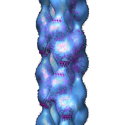
- Downloads & links
-EMDB archive
| Map data | emd_1313.map.gz | 609.9 KB | EMDB map data format | |
|---|---|---|---|---|
| Header (meta data) | emd-1313-v30.xmlemd-1313.xml | 10.3 KB 10.3 KB | Display Display | EMDB header |
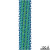
| Images |  1313.gif 1313.gif | 60.7 KB | ||
| Archive directory |  http://ftp.pdbj.org/pub/emdb/structures/EMD-1313ftp://ftp.pdbj.org/pub/emdb/structures/EMD-1313 http://ftp.pdbj.org/pub/emdb/structures/EMD-1313ftp://ftp.pdbj.org/pub/emdb/structures/EMD-1313 | HTTPS FTP |
-Related structure data
| Similar structure data |
|---|


-Links
| EMDB pages | EMDB (EBI/PDBe) / EMDataResource |
|---|
-Map
| File | Download / File: emd_1313.map.gz / Format: CCP4 / Size: 7.8 MB / Type: IMAGE STORED AS FLOATING POINT NUMBER (4 BYTES) | ||||||||||||||||||||||||||||||||||||||||||||||||||||||||||||||||||||
|---|---|---|---|---|---|---|---|---|---|---|---|---|---|---|---|---|---|---|---|---|---|---|---|---|---|---|---|---|---|---|---|---|---|---|---|---|---|---|---|---|---|---|---|---|---|---|---|---|---|---|---|---|---|---|---|---|---|---|---|---|---|---|---|---|---|---|---|---|---|
| Annotation | Map of the carboxy-terminal truncated nitrilase helix from Rhodococcus rhodochrous J1 | ||||||||||||||||||||||||||||||||||||||||||||||||||||||||||||||||||||
| Projections & slices | Image control
Images are generated by Spider. | ||||||||||||||||||||||||||||||||||||||||||||||||||||||||||||||||||||
| Voxel size | X=Y=Z: 4 Å | ||||||||||||||||||||||||||||||||||||||||||||||||||||||||||||||||||||
| Density |
| ||||||||||||||||||||||||||||||||||||||||||||||||||||||||||||||||||||
| Symmetry | Space group: 1 | ||||||||||||||||||||||||||||||||||||||||||||||||||||||||||||||||||||
| Details | EMDB XML:
CCP4 map header:
| ||||||||||||||||||||||||||||||||||||||||||||||||||||||||||||||||||||
 Z (Sec.)
 Y (Row.)
 X (Col.)


-Supplemental data
- Sample components
-Entire : Carboxy-terminal truncated nitrilase from Rhodococcus rhodochrous J1
| Entire | Name: Carboxy-terminal truncated nitrilase from Rhodococcus rhodochrous J1 |
|---|---|
| Components |
|
-Supramolecule #1000: Carboxy-terminal truncated nitrilase from Rhodococcus rhodochrous J1
| Supramolecule | Name: Carboxy-terminal truncated nitrilase from Rhodococcus rhodochrous J1 type: sample / ID: 1000 / Number unique components: 1 |
|---|---|
| Molecular weight | Experimental: 36 KDa / Theoretical: 36 KDa / Method: SDS-PAGE MALDI-TOF mass spectrometry |
-Macromolecule #1: J1 nitrilase 1-327
| Macromolecule | Name: J1 nitrilase 1-327 / type: protein_or_peptide / ID: 1 / Oligomeric state: helix / Recombinant expression: Yes |
|---|---|
| Source (natural) | Organism: Rhodococcus rhodochrous (bacteria) / Strain: J1 |
| Molecular weight | Experimental: 36 KDa / Theoretical: 36 KDa |
| Recombinant expression | Organism: |
| Sequence | GO: nitrilase activity / InterPro: Carbon-nitrogen hydrolase |
-Experimental details
-Structure determination
| Method | negative staining |
|---|---|
 Processing Processing | helical reconstruction |
| Aggregation state | filament |
-Sample preparation
| Concentration | 0.5 mg/mL |
|---|---|
| Buffer | pH: 7.8 / Details: 100mM KH2PO4, 400mM KCl, 10% (v/v) EtOH |
| Staining | Type: NEGATIVE Details: Sample was applied to glow discharged carbon films which were then washed twice with distilled water and then stained with 2% w/v uranyl acetate |
| Grid | Details: 300 mesh |
| Vitrification | Cryogen name: NONE |
- Electron microscopy
| Microscope | JEOL 2000EX |
|---|---|
| Image recording | Category: FILM / Film or detector model: KODAK SO-163 FILM / Digitization - Scanner: OTHER / Digitization - Sampling interval: 10 µm / Number real images: 50 / Average electron dose: 30 e/Å2 / Bits/pixel: 16 |
| Electron beam | Acceleration voltage: 120 kV / Electron source: TUNGSTEN HAIRPIN |
| Electron optics | Calibrated magnification: 50000 / Illumination mode: FLOOD BEAM / Imaging mode: BRIGHT FIELD / Nominal magnification: 50000 |
| Sample stage | Specimen holder: standard / Specimen holder model: OTHER |
-Image processing
| Details | Helices were formed by autolysis of the wild-type protein after storage for one month at 4 degrees C |
|---|---|
| Final reconstruction | Applied symmetry - Helical parameters - Δz: 15.8 Å Applied symmetry - Helical parameters - Δ&Phi: 73.65 ° Applied symmetry - Helical parameters - Axial symmetry: D1 (2x1 fold dihedral) Algorithm: OTHER / Resolution.type: BY AUTHOR / Resolution: 18.0 Å / Resolution method: FSC 0.5 CUT-OFF / Software - Name: SPIDER / Details: D1 symmetry was imposed on map after convergence |
-Atomic model buiding 1
| Software | Name: Situs |
|---|---|
| Details | The helical symmetry was applied to a homology model to generate a helix containing 9 dimers. This was fitted to the map using CoLoRes. A two dimensional search of radial distance and azimuthal angle was conducted to find the best fit. |
| Refinement | Space: REAL / Protocol: RIGID BODY FIT |
