National Institutes of Health/National Institute of General Medical Sciences (NIH/NIGMS)
R01 GM118396
United States
National Institutes of Health/National Cancer Institute (NIH/NCI)
CA197855
United States
National Institutes of Health/National Institute of General Medical Sciences (NIH/NIGMS)
R01 GM047414
United States
Citation
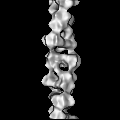
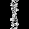
Journal: J Cell Biol / Year: 2017 Title: The glycolytic enzyme phosphofructokinase-1 assembles into filaments. Authors: Bradley A Webb / Anne M Dosey / Torsten Wittmann / Justin M Kollman / Diane L Barber / Abstract: Despite abundant knowledge of the regulation and biochemistry of glycolytic enzymes, we have limited understanding on how they are spatially organized in the cell. Emerging evidence indicates that ...Despite abundant knowledge of the regulation and biochemistry of glycolytic enzymes, we have limited understanding on how they are spatially organized in the cell. Emerging evidence indicates that nonglycolytic metabolic enzymes regulating diverse pathways can assemble into polymers. We now show tetramer- and substrate-dependent filament assembly by phosphofructokinase-1 (PFK1), which is considered the "gatekeeper" of glycolysis because it catalyzes the step committing glucose to breakdown. Recombinant liver PFK1 (PFKL) isoform, but not platelet PFK1 (PFKP) or muscle PFK1 (PFKM) isoforms, assembles into filaments. Negative-stain electron micrographs reveal that filaments are apolar and made of stacked tetramers oriented with exposed catalytic sites positioned along the edge of the polymer. Electron micrographs and biochemical data with a PFKL/PFKP chimera indicate that the PFKL regulatory domain mediates filament assembly. Quantified live-cell imaging shows dynamic properties of localized PFKL puncta that are enriched at the plasma membrane. These findings reveal a new behavior of a key glycolytic enzyme with insights on spatial organization and isoform-specific glucose metabolism in cells.
History
Deposition
Dec 27, 2016
-
Header (metadata) release
Jan 11, 2017
-
Map release
Jan 24, 2018
-
Update
Jan 29, 2020
-
Current status
Jan 29, 2020
Processing site: RCSB / Status: Released
-
Structure visualization
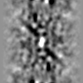
Movie
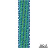
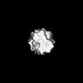
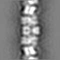
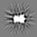
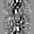
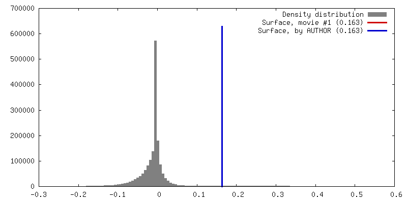
Surface view with section colored by density value
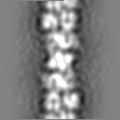
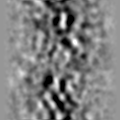
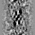
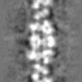
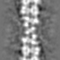
Entire : Filament of phosphofructokinase-1 liver isoform
Entire
Name: Filament of phosphofructokinase-1 liver isoform
Components
Complex: Filament of phosphofructokinase-1 liver isoform
-
Supramolecule #1: Filament of phosphofructokinase-1 liver isoform
Supramolecule
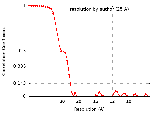
Name: Filament of phosphofructokinase-1 liver isoform / type: complex / ID: 1 / Parent: 0 Details: PFKL tetramers assembled into filaments by addition of ATP and fructose-6-phosphate
Material: COPPER / Mesh: 400 / Support film - Material: CARBON / Support film - topology: CONTINUOUS / Pretreatment - Type: GLOW DISCHARGE
-
Electron microscopy
Microscope
FEI TECNAI SPIRIT
Image recording
Film or detector model: GATAN ULTRASCAN 4000 (4k x 4k) / Digitization - Dimensions - Width: 4096 pixel / Digitization - Dimensions - Height: 4096 pixel / Number grids imaged: 1 / Number real images: 120 / Average exposure time: 1.2 sec. / Average electron dose: 30.0 e/Å2
Electron beam
Acceleration voltage: 120 kV / Electron source: LAB6
Electron optics
Illumination mode: FLOOD BEAM / Imaging mode: BRIGHT FIELD
Sample stage
Specimen holder model: SIDE ENTRY, EUCENTRIC
Experimental equipment
Model: Tecnai Spirit / Image courtesy: FEI Company
In the structure databanks used in Yorodumi, some data are registered as the other names, "COVID-19 virus" and "2019-nCoV". Here are the details of the virus and the list of structure data.
Jan 31, 2019. EMDB accession codes are about to change! (news from PDBe EMDB page)
EMDB accession codes are about to change! (news from PDBe EMDB page)
The allocation of 4 digits for EMDB accession codes will soon come to an end. Whilst these codes will remain in use, new EMDB accession codes will include an additional digit and will expand incrementally as the available range of codes is exhausted. The current 4-digit format prefixed with “EMD-” (i.e. EMD-XXXX) will advance to a 5-digit format (i.e. EMD-XXXXX), and so on. It is currently estimated that the 4-digit codes will be depleted around Spring 2019, at which point the 5-digit format will come into force.
The EM Navigator/Yorodumi systems omit the EMD- prefix.
Related info.:Q: What is EMD? / ID/Accession-code notation in Yorodumi/EM Navigator
Yorodumi is a browser for structure data from EMDB, PDB, SASBDB, etc.
This page is also the successor to EM Navigator detail page, and also detail information page/front-end page for Omokage search.
The word "yorodu" (or yorozu) is an old Japanese word meaning "ten thousand". "mi" (miru) is to see.
Related info.:EMDB / PDB / SASBDB / Comparison of 3 databanks / Yorodumi Search / Aug 31, 2016. New EM Navigator & Yorodumi / Yorodumi Papers / Jmol/JSmol / Function and homology information / Changes in new EM Navigator and Yorodumi
 Movie
Movie Controller


 Open data
 Basic information
 Map data
 Sample
 Authors
 United States, 3 items
  Citation
 Structure visualization
 Movie viewer
 UCSF Chimera
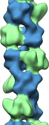
 Downloads & links
 emd_8542.png
 http://ftp.pdbj.org/pub/emdb/structures/EMD-8542
 Z (Sec.)
 Y (Row.)
 X (Col.)


 Sample components
 Spodoptera frugiperda (fall armyworm) / Recombinant plasmid: pFastBac
 Processing
 Electron microscopy