- EMDB-3581: Structure of 70S ribosome from Lactococcus lactis -
+
Open data
ID or keywords:
Loading...
-
Basic information
Entry
Database: EMDB / ID: EMD-3581
Title
Structure of 70S ribosome from Lactococcus lactis
Map data
Sample
Complex: 70S ribosome
RNA: x 3 types
Protein or peptide: x 48 types
Keywords
ribosome / 70S / lactoccocus lactis / Cryo-EM
Function / homology
Function and homology information
negative regulation of translational elongation / ribosomal small subunit binding / large ribosomal subunit / transferase activity / ribosomal small subunit assembly / ribosome biogenesis / ribosomal small subunit biogenesis / 5S rRNA binding / ribosomal large subunit assembly / small ribosomal subunit ...negative regulation of translational elongation / ribosomal small subunit binding / large ribosomal subunit / transferase activity / ribosomal small subunit assembly / ribosome biogenesis / ribosomal small subunit biogenesis / 5S rRNA binding / ribosomal large subunit assembly / small ribosomal subunit / small ribosomal subunit rRNA binding / large ribosomal subunit rRNA binding / cytosolic small ribosomal subunit / cytosolic large ribosomal subunit / cytoplasmic translation / tRNA binding / negative regulation of translation / rRNA binding / structural constituent of ribosome / ribosome / translation / ribonucleoprotein complex / mRNA binding / RNA binding / zinc ion binding / cytoplasm / cytosol Similarity search - Function
Ribosome hibernation promoting factor, long/plastid / Sigma 54 modulation/S30EA ribosomal protein, C-terminal / Sigma 54 modulation/S30EA ribosomal protein, C-terminal domain superfamily / Sigma 54 modulation/S30EA ribosomal protein C terminus / : / Ribosome hibernation promoting factor/RaiA / Ribosome hibernation promotion factor-like / Sigma 54 modulation protein / S30EA ribosomal protein / Ribosomal protein L31 type B / : ...Ribosome hibernation promoting factor, long/plastid / Sigma 54 modulation/S30EA ribosomal protein, C-terminal / Sigma 54 modulation/S30EA ribosomal protein, C-terminal domain superfamily / Sigma 54 modulation/S30EA ribosomal protein C terminus / : / Ribosome hibernation promoting factor/RaiA / Ribosome hibernation promotion factor-like / Sigma 54 modulation protein / S30EA ribosomal protein / Ribosomal protein L31 type B / : / Ribosomal protein S14, type Z / Ribosomal protein S21, conserved site / Ribosomal protein S21 signature. / Ribosomal protein S21 superfamily / Ribosomal protein S21 / Ribosomal protein L31 signature. / Ribosomal protein L31 / Ribosomal protein L31 superfamily / Ribosomal protein L31 / Ribosomal protein S21 / Ribosomal protein L16 signature 1. / Ribosomal protein L6, conserved site / Ribosomal protein L6 signature 1. / : / Ribosomal protein L16 signature 2. / Ribosomal protein L16, conserved site / Ribosomal protein L17 signature. / Ribosomal protein L36 signature. / : / Ribosomal protein L28/L24 superfamily / Ribosomal protein L32p, bacterial type / Ribosomal protein L35, conserved site / Ribosomal protein L35 signature. / Ribosomal protein L28 / Ribosomal protein L35, non-mitochondrial / Ribosomal protein L18, bacterial-type / Ribosomal protein S3, bacterial-type / Ribosomal protein S13, bacterial-type / Ribosomal protein S19, bacterial-type / : / Ribosomal protein L6, bacterial-type / Ribosomal protein S7, bacterial/organellar-type / Ribosomal protein S11, bacterial-type / Ribosomal protein S20 / Ribosomal protein S20 superfamily / Ribosomal protein S20 / Ribosomal protein S4, bacterial-type / Ribosomal protein L5, bacterial-type / Ribosomal protein S5, bacterial-type / Ribosomal protein L19, conserved site / Ribosomal protein L19 signature. / 30S ribosomal protein S17 / : / Ribosomal protein S6, plastid/chloroplast / Ribosomal protein L36 / Ribosomal protein L36 superfamily / Ribosomal protein L36 / Ribosomal protein L20 signature. / Ribosomal protein L34, conserved site / Ribosomal protein L34 signature. / Ribosomal protein S14/S29 / Ribosomal protein L14P, bacterial-type / Ribosomal protein L27, conserved site / Ribosomal protein L27 signature. / Ribosomal protein S2, bacteria/mitochondria/plastid / Ribosomal protein L35 / Ribosomal protein L35 superfamily / Ribosomal protein L22, bacterial/chloroplast-type / Ribosomal protein L35 / Ribosomal protein L2, bacterial/organellar-type / Ribosomal protein L33 / Ribosomal protein L18 / Ribosomal L18 of archaea, bacteria, mitoch. and chloroplast / Ribosomal protein S18, conserved site / Ribosomal protein S18 signature. / Ribosomal protein L33 / Ribosomal L28 family / Ribosomal protein S9, bacterial/plastid / Ribosomal protein L33 superfamily / Ribosomal protein L28/L24 / Ribosomal protein L30, bacterial-type / Ribosomal protein S16 / Ribosomal protein S16 domain superfamily / Ribosomal protein S16 / L28p-like / Ribosomal protein L16 / Ribosomal protein S15, bacterial-type / Ribosomal protein S6 / Ribosomal protein S6 / Ribosomal protein S6 superfamily / Ribosomal protein L20 / Ribosomal protein S12, bacterial-type / Ribosomal protein L20 / Ribosomal protein L20, C-terminal / Ribosomal protein L19 / Ribosomal protein L19 / Ribosomal protein L19 superfamily / : / Large ribosomal subunit protein uL24, C-terminal domain / Translation elongation factor EF1B/ribosomal protein S6 Similarity search - Domain/homology
Large ribosomal subunit protein bL32 / Large ribosomal subunit protein bL34 / Large ribosomal subunit protein bL28 / Small ribosomal subunit protein bS21 / Small ribosomal subunit protein uS4 / Ribosome hibernation promotion factor / Large ribosomal subunit protein bL31B / Small ribosomal subunit protein bS16 / Large ribosomal subunit protein bL27 / Large ribosomal subunit protein bL21 ...Large ribosomal subunit protein bL32 / Large ribosomal subunit protein bL34 / Large ribosomal subunit protein bL28 / Small ribosomal subunit protein bS21 / Small ribosomal subunit protein uS4 / Ribosome hibernation promotion factor / Large ribosomal subunit protein bL31B / Small ribosomal subunit protein bS16 / Large ribosomal subunit protein bL27 / Large ribosomal subunit protein bL21 / Large ribosomal subunit protein bL19 / Small ribosomal subunit protein bS20 / Large ribosomal subunit protein bL20 / Large ribosomal subunit protein bL35 / Small ribosomal subunit protein uS15 / Large ribosomal subunit protein bL17 / Small ribosomal subunit protein uS11 / Small ribosomal subunit protein uS13 / Large ribosomal subunit protein bL36 / Large ribosomal subunit protein uL15 / Large ribosomal subunit protein uL30 / Small ribosomal subunit protein uS5 / Large ribosomal subunit protein uL18 / Large ribosomal subunit protein uL6 / Small ribosomal subunit protein uS8 / Small ribosomal subunit protein uS14 / Large ribosomal subunit protein uL5 / Large ribosomal subunit protein uL24 / Large ribosomal subunit protein uL14 / Small ribosomal subunit protein uS17 / Large ribosomal subunit protein uL29 / Large ribosomal subunit protein uL16 / Small ribosomal subunit protein uS3 / Large ribosomal subunit protein uL22 / Small ribosomal subunit protein uS19 / Large ribosomal subunit protein uL2 / Large ribosomal subunit protein uL23 / Large ribosomal subunit protein uL4 / Large ribosomal subunit protein uL3 / Small ribosomal subunit protein uS10 / Large ribosomal subunit protein bL33C / Small ribosomal subunit protein uS2 / Small ribosomal subunit protein bS18 / Small ribosomal subunit protein bS6 / Small ribosomal subunit protein uS9 / Large ribosomal subunit protein uL13 / Small ribosomal subunit protein uS7 / Small ribosomal subunit protein uS12 Similarity search - Component
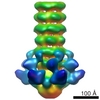
Journal: Nat Commun / Year: 2017 Title: A general mechanism of ribosome dimerization revealed by single-particle cryo-electron microscopy. Authors: Linda E Franken / Gert T Oostergetel / Tjaard Pijning / Pranav Puri / Valentina Arkhipova / Egbert J Boekema / Bert Poolman / Albert Guskov / Abstract: Bacteria downregulate their ribosomal activity through dimerization of 70S ribosomes, yielding inactive 100S complexes. In Escherichia coli, dimerization is mediated by the hibernation promotion ...Bacteria downregulate their ribosomal activity through dimerization of 70S ribosomes, yielding inactive 100S complexes. In Escherichia coli, dimerization is mediated by the hibernation promotion factor (HPF) and ribosome modulation factor. Here we report the cryo-electron microscopy study on 100S ribosomes from Lactococcus lactis and a dimerization mechanism involving a single protein: HPF. The N-terminal domain of HPF binds at the same site as HPF in Escherichia coli 100S ribosomes. Contrary to ribosome modulation factor, the C-terminal domain of HPF binds exactly at the dimer interface. Furthermore, ribosomes from Lactococcus lactis do not undergo conformational changes in the 30S head domains upon binding of HPF, and the Shine-Dalgarno sequence and mRNA entrance tunnel remain accessible. Ribosome activity is blocked by HPF due to the inhibition of mRNA recognition by the platform binding center. Phylogenetic analysis of HPF proteins suggests that HPF-mediated dimerization is a widespread mechanism of ribosome hibernation in bacteria.When bacteria enter the stationary growth phase, protein translation is suppressed via the dimerization of 70S ribosomes into inactive complexes. Here the authors provide a structural basis for how the dual domain hibernation promotion factor promotes ribosome dimerization and hibernation in bacteria.
History
Deposition
Jan 26, 2017
-
Header (metadata) release
Feb 15, 2017
-
Map release
Oct 11, 2017
-
Update
May 15, 2024
-
Current status
May 15, 2024
Processing site: PDBe / Status: Released
-
Structure visualization
Movie
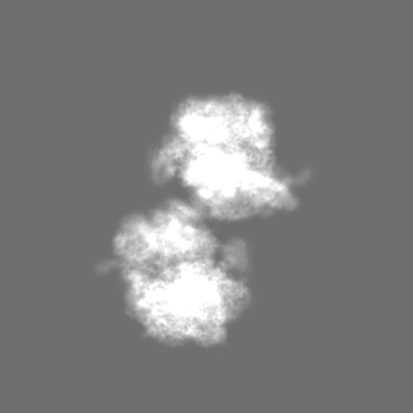
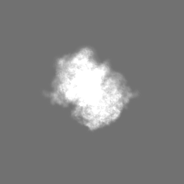
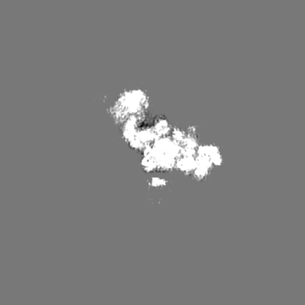
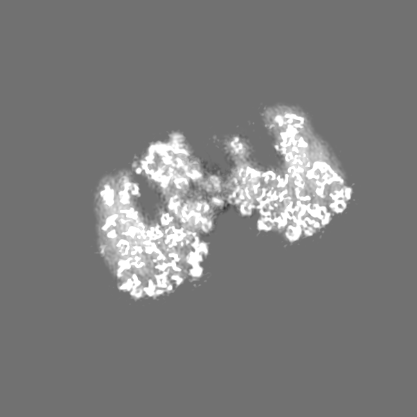
Surface view with section colored by density value
In the structure databanks used in Yorodumi, some data are registered as the other names, "COVID-19 virus" and "2019-nCoV". Here are the details of the virus and the list of structure data.
Jan 31, 2019. EMDB accession codes are about to change! (news from PDBe EMDB page)
EMDB accession codes are about to change! (news from PDBe EMDB page)
The allocation of 4 digits for EMDB accession codes will soon come to an end. Whilst these codes will remain in use, new EMDB accession codes will include an additional digit and will expand incrementally as the available range of codes is exhausted. The current 4-digit format prefixed with “EMD-” (i.e. EMD-XXXX) will advance to a 5-digit format (i.e. EMD-XXXXX), and so on. It is currently estimated that the 4-digit codes will be depleted around Spring 2019, at which point the 5-digit format will come into force.
The EM Navigator/Yorodumi systems omit the EMD- prefix.
Related info.:Q: What is EMD? / ID/Accession-code notation in Yorodumi/EM Navigator
Yorodumi is a browser for structure data from EMDB, PDB, SASBDB, etc.
This page is also the successor to EM Navigator detail page, and also detail information page/front-end page for Omokage search.
The word "yorodu" (or yorozu) is an old Japanese word meaning "ten thousand". "mi" (miru) is to see.
Related info.:EMDB / PDB / SASBDB / Comparison of 3 databanks / Yorodumi Search / Aug 31, 2016. New EM Navigator & Yorodumi / Yorodumi Papers / Jmol/JSmol / Function and homology information / Changes in new EM Navigator and Yorodumi
 Movie
Movie Controller


 Open data
 Basic information
 Map data
 Sample
 Keywords
 Function and homology information
 Lactococcus lactis (lactic acid bacteria) /
  Authors
 Netherlands, 2 items
  Citation
 Structure visualization
 Downloads & links
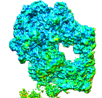 emd_3581.png
 http://ftp.pdbj.org/pub/emdb/structures/EMD-3581


 Z (Sec.)
 Y (Row.)
 X (Col.)


 Sample components
 Processing
 Electron microscopy
 FIELD EMISSION GUN