 Movie
Movie Controller


+ Open data

- Basic information
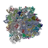
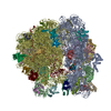
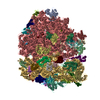
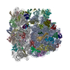
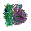
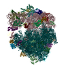
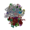
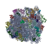
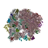
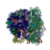
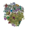
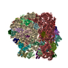
| Entry | Database: PDB / ID: 5myj | |||||||||
|---|---|---|---|---|---|---|---|---|---|---|
| Title | Structure of 70S ribosome from Lactococcus lactis | |||||||||
 Components Components |
| |||||||||
 Keywords Keywords | RIBOSOME / 70S / lactoccocus lactis / Cryo-EM | |||||||||
| Function / homology |  Function and homology information Function and homology informationnegative regulation of translational elongation / ribosomal small subunit binding / large ribosomal subunit / transferase activity / ribosomal small subunit assembly / ribosome biogenesis / ribosomal small subunit biogenesis / 5S rRNA binding / ribosomal large subunit assembly / small ribosomal subunit ...negative regulation of translational elongation / ribosomal small subunit binding / large ribosomal subunit / transferase activity / ribosomal small subunit assembly / ribosome biogenesis / ribosomal small subunit biogenesis / 5S rRNA binding / ribosomal large subunit assembly / small ribosomal subunit / small ribosomal subunit rRNA binding / large ribosomal subunit rRNA binding / cytosolic small ribosomal subunit / cytosolic large ribosomal subunit / cytoplasmic translation / tRNA binding / negative regulation of translation / rRNA binding / structural constituent of ribosome / ribosome / translation / ribonucleoprotein complex / mRNA binding / RNA binding / zinc ion binding / cytoplasm / cytosol Similarity search - Function | |||||||||
| Biological species |  Lactococcus lactis subsp. cremoris MG1363 (lactic acid bacteria)Lactococcus lactis subsp. cremoris (lactic acid bacteria) Lactococcus lactis subsp. cremoris MG1363 (lactic acid bacteria)Lactococcus lactis subsp. cremoris (lactic acid bacteria) | |||||||||
| Method | ELECTRON MICROSCOPY / single particle reconstruction / cryo EM / Resolution: 5.6 Å | |||||||||
 Authors Authors | Franken, L.E. / Oostergetel, G.T. / Pijning, T. / Puri, P. / Boekema, E.J. / Poolman, B. / Guskov, A. | |||||||||
| Funding support |  Netherlands, 2items Netherlands, 2items
| |||||||||
 Citation Citation | Journal: Nat Commun / Year: 2017 Title: A general mechanism of ribosome dimerization revealed by single-particle cryo-electron microscopy. Authors: Linda E Franken / Gert T Oostergetel / Tjaard Pijning / Pranav Puri / Valentina Arkhipova / Egbert J Boekema / Bert Poolman / Albert Guskov / Abstract: Bacteria downregulate their ribosomal activity through dimerization of 70S ribosomes, yielding inactive 100S complexes. In Escherichia coli, dimerization is mediated by the hibernation promotion ...Bacteria downregulate their ribosomal activity through dimerization of 70S ribosomes, yielding inactive 100S complexes. In Escherichia coli, dimerization is mediated by the hibernation promotion factor (HPF) and ribosome modulation factor. Here we report the cryo-electron microscopy study on 100S ribosomes from Lactococcus lactis and a dimerization mechanism involving a single protein: HPF. The N-terminal domain of HPF binds at the same site as HPF in Escherichia coli 100S ribosomes. Contrary to ribosome modulation factor, the C-terminal domain of HPF binds exactly at the dimer interface. Furthermore, ribosomes from Lactococcus lactis do not undergo conformational changes in the 30S head domains upon binding of HPF, and the Shine-Dalgarno sequence and mRNA entrance tunnel remain accessible. Ribosome activity is blocked by HPF due to the inhibition of mRNA recognition by the platform binding center. Phylogenetic analysis of HPF proteins suggests that HPF-mediated dimerization is a widespread mechanism of ribosome hibernation in bacteria.When bacteria enter the stationary growth phase, protein translation is suppressed via the dimerization of 70S ribosomes into inactive complexes. Here the authors provide a structural basis for how the dual domain hibernation promotion factor promotes ribosome dimerization and hibernation in bacteria. | |||||||||
| History |
|
- Structure visualization
| Movie |
Movie viewer |
|---|---|
| Structure viewer | Molecule: MolmilJmol/JSmol |
- Downloads & links
-Download
| PDBx/mmCIF format | 5myj.cif.gz | 3.1 MB | Display | PDBx/mmCIF format |
|---|---|---|---|---|
| PDB format | pdb5myj.ent.gz | Display | PDB format | |
| PDBx/mmJSON format | 5myj.json.gz | Tree view | PDBx/mmJSON format | |
| Others |  Other downloads Other downloads |
-Validation report
| Arichive directory | https://data.pdbj.org/pub/pdb/validation_reports/my/5myjftp://data.pdbj.org/pub/pdb/validation_reports/my/5myj | HTTPS FTP |
|---|
-Related structure data
| Related structure data |  3581MC M: map data used to model this data C: citing same article ( |
|---|---|
| Similar structure data |


-Links
 PDBj
PDBj


- Assembly
| Deposited unit | 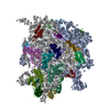
|
|---|---|
| 1 |
|
-Components
-RNA chain , 3 types, 3 molecules AABABB
| #1: RNA chain | Mass: 497142.344 Da / Num. of mol.: 1 / Source method: isolated from a natural source Source: (natural) Lactococcus lactis subsp. cremoris MG1363 (lactic acid bacteria)References: GenBank: 124491690 |
|---|---|
| #31: RNA chain | Mass: 938591.125 Da / Num. of mol.: 1 / Source method: isolated from a natural source Source: (natural) Lactococcus lactis subsp. cremoris MG1363 (lactic acid bacteria)References: GenBank: 124491690 |
| #32: RNA chain | Mass: 37065.969 Da / Num. of mol.: 1 / Source method: isolated from a natural source Source: (natural) Lactococcus lactis subsp. cremoris MG1363 (lactic acid bacteria)References: GenBank: 124491690 |
-30S ribosomal protein ... , 20 types, 20 molecules ABACADAEAFAGAHAIAJAKALAMANAOAPAQARASATAU
| #2: Protein | Mass: 28589.760 Da / Num. of mol.: 1 / Source method: isolated from a natural source Source: (natural) Lactococcus lactis subsp. cremoris (strain MG1363) (lactic acid bacteria)Strain: MG1363 / References: UniProt: A2RNV0 |
|---|---|
| #3: Protein | Mass: 24058.795 Da / Num. of mol.: 1 / Source method: isolated from a natural source Source: (natural) Lactococcus lactis subsp. cremoris (strain MG1363) (lactic acid bacteria)Strain: MG1363 / References: UniProt: A2RNP9 |
| #4: Protein | Mass: 23232.605 Da / Num. of mol.: 1 / Source method: isolated from a natural source Source: (natural) Lactococcus lactis subsp. cremoris (strain MG1363) (lactic acid bacteria)Strain: MG1363 / References: UniProt: A2RI10 |
| #5: Protein | Mass: 17609.285 Da / Num. of mol.: 1 / Source method: isolated from a natural source Source: (natural) Lactococcus lactis subsp. cremoris (strain MG1363) (lactic acid bacteria)Strain: MG1363 / References: UniProt: A2RNN6 |
| #6: Protein | Mass: 11321.831 Da / Num. of mol.: 1 / Source method: isolated from a natural source Source: (natural) Lactococcus lactis subsp. cremoris (strain MG1363) (lactic acid bacteria)Strain: MG1363 / References: UniProt: A2RNZ4 |
| #7: Protein | Mass: 17713.443 Da / Num. of mol.: 1 / Source method: isolated from a natural source Source: (natural) Lactococcus lactis subsp. cremoris (strain MG1363) (lactic acid bacteria)Strain: MG1363 / References: UniProt: A2RP73 |
| #8: Protein | Mass: 14692.106 Da / Num. of mol.: 1 / Source method: isolated from a natural source Source: (natural) Lactococcus lactis subsp. cremoris (strain MG1363) (lactic acid bacteria)Strain: MG1363 / References: UniProt: A2RNN9 |
| #9: Protein | Mass: 14124.289 Da / Num. of mol.: 1 / Source method: isolated from a natural source Source: (natural) Lactococcus lactis subsp. cremoris (strain MG1363) (lactic acid bacteria)Strain: MG1363 / References: UniProt: A2RP61 |
| #10: Protein | Mass: 11764.765 Da / Num. of mol.: 1 / Source method: isolated from a natural source Source: (natural) Lactococcus lactis subsp. cremoris (strain MG1363) (lactic acid bacteria)Strain: MG1363 / References: UniProt: A2RNQ6 |
| #11: Protein | Mass: 13310.352 Da / Num. of mol.: 1 / Source method: isolated from a natural source Source: (natural) Lactococcus lactis subsp. cremoris (strain MG1363) (lactic acid bacteria)Strain: MG1363 / References: UniProt: A2RNM8 |
| #12: Protein | Mass: 15157.884 Da / Num. of mol.: 1 / Source method: isolated from a natural source Source: (natural) Lactococcus lactis subsp. cremoris (strain MG1363) (lactic acid bacteria)Strain: MG1363 / References: UniProt: A2RP74 |
| #13: Protein | Mass: 13519.672 Da / Num. of mol.: 1 / Source method: isolated from a natural source Source: (natural) Lactococcus lactis subsp. cremoris (strain MG1363) (lactic acid bacteria)Strain: MG1363 / References: UniProt: A2RNM9 |
| #14: Protein | Mass: 7151.611 Da / Num. of mol.: 1 / Source method: isolated from a natural source Source: (natural) Lactococcus lactis subsp. cremoris (strain MG1363) (lactic acid bacteria)Strain: MG1363 / References: UniProt: A2RNP2 |
| #15: Protein | Mass: 10367.041 Da / Num. of mol.: 1 / Source method: isolated from a natural source Source: (natural) Lactococcus lactis subsp. cremoris (strain MG1363) (lactic acid bacteria)Strain: MG1363 / References: UniProt: A2RMV9 |
| #16: Protein | Mass: 10270.938 Da / Num. of mol.: 1 / Source method: isolated from a natural source Source: (natural) Lactococcus lactis subsp. cremoris (strain MG1363) (lactic acid bacteria)Strain: MG1363 / References: UniProt: A2RJS1 |
| #17: Protein | Mass: 10166.831 Da / Num. of mol.: 1 / Source method: isolated from a natural source Source: (natural) Lactococcus lactis subsp. cremoris (strain MG1363) (lactic acid bacteria)Strain: MG1363 / References: UniProt: A2RNP6 |
| #18: Protein | Mass: 9392.087 Da / Num. of mol.: 1 / Source method: isolated from a natural source Source: (natural) Lactococcus lactis subsp. cremoris (strain MG1363) (lactic acid bacteria)Strain: MG1363 / References: UniProt: A2RNZ2 |
| #19: Protein | Mass: 10595.238 Da / Num. of mol.: 1 / Source method: isolated from a natural source Source: (natural) Lactococcus lactis subsp. cremoris (strain MG1363) (lactic acid bacteria)Strain: MG1363 / References: UniProt: A2RNQ1 |
| #20: Protein | Mass: 8384.740 Da / Num. of mol.: 1 / Source method: isolated from a natural source Source: (natural) Lactococcus lactis subsp. cremoris (strain MG1363) (lactic acid bacteria)Strain: MG1363 / References: UniProt: A2RMG0 |
| #21: Protein | Mass: 7041.308 Da / Num. of mol.: 1 / Source method: isolated from a natural source Source: (natural) Lactococcus lactis subsp. cremoris (strain MG1363) (lactic acid bacteria)Strain: MG1363 / References: UniProt: A2RHW9 |
+50S ribosomal protein ... , 27 types, 27 molecules B0B1B2B3B4B5B6B7B8BDBEBFBGBHBMBNBOBPBQBRBSBTBUBVBWBXBZ
-Protein , 1 types, 1 molecules A
| #51: Protein | Mass: 21345.988 Da / Num. of mol.: 1 / Source method: isolated from a natural source Source: (natural) Lactococcus lactis subsp. cremoris (strain MG1363) (lactic acid bacteria)Strain: MG1363 / References: UniProt: A2RIX0 |
|---|
-Experimental details
-Experiment
| Experiment | Method: ELECTRON MICROSCOPY |
|---|---|
| EM experiment | Aggregation state: PARTICLE / 3D reconstruction method: single particle reconstruction |
- Sample preparation
| Component | Name: 70S ribosome / Type: RIBOSOME / Entity ID: all / Source: NATURAL |
|---|---|
| Molecular weight | Experimental value: NO |
| Source (natural) | Organism: Lactococcus lactis (lactic acid bacteria) |
| Buffer solution | pH: 7.6 |
| Specimen | Embedding applied: NO / Shadowing applied: NO / Staining applied: NO / Vitrification applied: YES |
| Specimen support | Grid material: COPPER / Grid mesh size: 400 divisions/in. / Grid type: Quantifoil R2/2 |
| Vitrification | Instrument: FEI VITROBOT MARK III / Cryogen name: ETHANE / Humidity: 80 % / Chamber temperature: 291.15 K |
- Electron microscopy imaging
| Experimental equipment |  Model: Titan Krios / Image courtesy: FEI Company |
|---|---|
| Microscopy | Model: FEI TITAN KRIOS |
| Electron gun | Electron source:  FIELD EMISSION GUN / Accelerating voltage: 300 kV / Illumination mode: FLOOD BEAM FIELD EMISSION GUN / Accelerating voltage: 300 kV / Illumination mode: FLOOD BEAM |
| Electron lens | Mode: BRIGHT FIELD |
| Image recording | Electron dose: 25 e/Å2 / Film or detector model: FEI FALCON II (4k x 4k) |
| EM imaging optics | Spherical aberration corrector: Cs-corrector (CEOS, Heidelberg, Germany) |
- Processing
| EM software |
| ||||||||||||||||||||||||||||||||||||
|---|---|---|---|---|---|---|---|---|---|---|---|---|---|---|---|---|---|---|---|---|---|---|---|---|---|---|---|---|---|---|---|---|---|---|---|---|---|
| CTF correction | Type: PHASE FLIPPING AND AMPLITUDE CORRECTION | ||||||||||||||||||||||||||||||||||||
| Symmetry | Point symmetry: C1 (asymmetric) | ||||||||||||||||||||||||||||||||||||
| 3D reconstruction | Resolution: 5.6 Å / Resolution method: FSC 0.143 CUT-OFF / Num. of particles: 43530 / Num. of class averages: 2 / Symmetry type: POINT | ||||||||||||||||||||||||||||||||||||
| Atomic model building | Protocol: FLEXIBLE FIT / Space: REAL | ||||||||||||||||||||||||||||||||||||
| Refinement | Highest resolution: 5.6 Å |
