 Movie
Movie Controller

[English] 日本語
 Yorodumi
- EMDB-1100: Structural insights into the assembly of the type III secretion n... -
+ Open data

- Basic information
| Entry | Database: EMDB / ID: EMD-1100 | |||||||||
|---|---|---|---|---|---|---|---|---|---|---|
| Title | Structural insights into the assembly of the type III secretion needle complex. | |||||||||
 Map data Map data | none | |||||||||
 Sample Sample |
| |||||||||
| Biological species |  Salmonella enterica subsp. enterica serovar Typhimurium (bacteria) / Salmonella tyhpimurium Salmonella enterica subsp. enterica serovar Typhimurium (bacteria) / Salmonella tyhpimurium | |||||||||
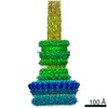
| Method | single particle reconstruction / cryo EM / Resolution: 17.0 Å | |||||||||
 Authors Authors | Marlovits TC / Kubori T / Sukhan A / Thomas DR / Galan JE / Unger VM | |||||||||
 Citation Citation | Journal: Science / Year: 2004 Title: Structural insights into the assembly of the type III secretion needle complex. Authors: Thomas C Marlovits / Tomoko Kubori / Anand Sukhan / Dennis R Thomas / Jorge E Galán / Vinzenz M Unger /  Abstract: Type III secretion systems (TTSSs) mediate translocation of virulence factors into host cells. We report the 17-angstrom resolution structures of a central component of Salmonella typhimurium TTSS, ...Type III secretion systems (TTSSs) mediate translocation of virulence factors into host cells. We report the 17-angstrom resolution structures of a central component of Salmonella typhimurium TTSS, the needle complex, and its assembly precursor, the bacterial envelope-anchored base. Both the base and the fully assembled needle complex adopted multiple oligomeric states in vivo, and needle assembly was accompanied by recruitment of the protein PrgJ as a structural component of the base. Moreover, conformational changes during needle assembly created scaffolds for anchoring both PrgJ and the needle substructure and may provide the basis for substrate-specificity switching during type III secretion. | |||||||||
| History |
|
- Structure visualization
| Movie |
 Movie viewer Movie viewer |
|---|---|
| Structure viewer | EM map: SurfViewMolmilJmol/JSmol |
| Supplemental images |

- Downloads & links
-EMDB archive
| Map data | emd_1100.map.gz | 24.8 MB | EMDB map data format | |
|---|---|---|---|---|
| Header (meta data) | emd-1100-v30.xmlemd-1100.xml | 10.9 KB 10.9 KB | Display Display | EMDB header |
| Images | 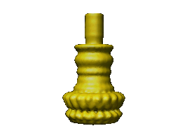 1100.gif 1100.gif | 9.3 KB | ||
| Archive directory |  http://ftp.pdbj.org/pub/emdb/structures/EMD-1100ftp://ftp.pdbj.org/pub/emdb/structures/EMD-1100 http://ftp.pdbj.org/pub/emdb/structures/EMD-1100ftp://ftp.pdbj.org/pub/emdb/structures/EMD-1100 | HTTPS FTP |
-Related structure data


-Links
| EMDB pages | EMDB (EBI/PDBe) / EMDataResource |
|---|
-Map
| File | Download / File: emd_1100.map.gz / Format: CCP4 / Size: 29.8 MB / Type: IMAGE STORED AS FLOATING POINT NUMBER (4 BYTES) | ||||||||||||||||||||||||||||||||||||||||||||||||||||||||||||||||||||
|---|---|---|---|---|---|---|---|---|---|---|---|---|---|---|---|---|---|---|---|---|---|---|---|---|---|---|---|---|---|---|---|---|---|---|---|---|---|---|---|---|---|---|---|---|---|---|---|---|---|---|---|---|---|---|---|---|---|---|---|---|---|---|---|---|---|---|---|---|---|
| Annotation | none | ||||||||||||||||||||||||||||||||||||||||||||||||||||||||||||||||||||
| Projections & slices | Image control
Images are generated by Spider. | ||||||||||||||||||||||||||||||||||||||||||||||||||||||||||||||||||||
| Voxel size | X=Y=Z: 2.8 Å | ||||||||||||||||||||||||||||||||||||||||||||||||||||||||||||||||||||
| Density |
| ||||||||||||||||||||||||||||||||||||||||||||||||||||||||||||||||||||
| Symmetry | Space group: 1 | ||||||||||||||||||||||||||||||||||||||||||||||||||||||||||||||||||||
| Details | EMDB XML:
CCP4 map header:
| ||||||||||||||||||||||||||||||||||||||||||||||||||||||||||||||||||||
 Z (Sec.)
 Y (Row.)
 X (Col.)


-Supplemental data
- Sample components
-Entire : Needle Complex of the Type III Secretion from Salmonella typhimurium
| Entire | Name: Needle Complex of the Type III Secretion from Salmonella typhimurium |
|---|---|
| Components |
|
-Supramolecule #1000: Needle Complex of the Type III Secretion from Salmonella typhimurium
| Supramolecule | Name: Needle Complex of the Type III Secretion from Salmonella typhimurium type: sample / ID: 1000 Details: theoretical molecular weight for the base (without the needle filament and the inner rod) is approximately 2.7MDa Oligomeric state: 20 / Number unique components: 5 |
|---|
-Macromolecule #1: PrgH
| Macromolecule | Name: PrgH / type: protein_or_peptide / ID: 1 / Recombinant expression: No |
|---|---|
| Source (natural) | Organism: Salmonella enterica subsp. enterica serovar Typhimurium (bacteria) |
-Macromolecule #2: PrgK
| Macromolecule | Name: PrgK / type: protein_or_peptide / ID: 2 / Recombinant expression: No |
|---|---|
| Source (natural) | Organism: Salmonella tyhpimurium |
| Molecular weight | Experimental: 28 MDa |
-Macromolecule #3: InvG
| Macromolecule | Name: InvG / type: protein_or_peptide / ID: 3 / Recombinant expression: No |
|---|---|
| Source (natural) | Organism: Salmonella enterica subsp. enterica serovar Typhimurium (bacteria) |
| Molecular weight | Experimental: 60 MDa |
-Macromolecule #4: PrgJ
| Macromolecule | Name: PrgJ / type: protein_or_peptide / ID: 4 / Recombinant expression: No |
|---|---|
| Source (natural) | Organism: Salmonella enterica subsp. enterica serovar Typhimurium (bacteria) |
| Molecular weight | Experimental: 10 MDa |
-Macromolecule #5: PRGI
| Macromolecule | Name: PRGI / type: protein_or_peptide / ID: 5 / Recombinant expression: No |
|---|---|
| Source (natural) | Organism: Salmonella tyhpimurium |
| Molecular weight | Experimental: 8 MDa |
-Experimental details
-Structure determination
| Method | cryo EM |
|---|---|
 Processing Processing | single particle reconstruction |
| Aggregation state | particle |
-Sample preparation
| Buffer | pH: 8 / Details: 10mM Tris-HCl 500mM NaCl 0.2% LDAO |
|---|---|
| Grid | Details: 400 mesh Cu/Rh grid |
| Vitrification | Cryogen name: ETHANE / Details: Vitrification carried out in the cold room / Method: Blot for 15 seconds before plunging |
- Electron microscopy
| Microscope | FEI TECNAI F20 |
|---|---|
| Temperature | Average: 95 K |
| Alignment procedure | Legacy - Astigmatism: 220,000 |
| Image recording | Category: FILM / Film or detector model: KODAK SO-163 FILM / Digitization - Scanner: ZEISS SCAI / Digitization - Sampling interval: 7 µm / Number real images: 79 / Average electron dose: 10 e/Å2 / Od range: 0.8 / Bits/pixel: 8 |
| Electron beam | Acceleration voltage: 200 kV / Electron source:  FIELD EMISSION GUN FIELD EMISSION GUN |
| Electron optics | Illumination mode: FLOOD BEAM / Imaging mode: BRIGHT FIELD / Cs: 2.0 mm / Nominal defocus max: 2.7 µm / Nominal defocus min: 1.3 µm / Nominal magnification: 50000 |
| Sample stage | Specimen holder: Side entry liquid nitrogen-cooled cryo specimen holder Specimen holder model: GATAN LIQUID NITROGEN |
| Experimental equipment |  Model: Tecnai F20 / Image courtesy: FEI Company |
-Image processing
| Details | The particles were selected interactively at the computer terminal. |
|---|---|
| CTF correction | Details: phase flipping, each particle |
| Final reconstruction | Applied symmetry - Point group: C20 (20 fold cyclic) / Algorithm: OTHER / Resolution.type: BY AUTHOR / Resolution: 17.0 Å / Resolution method: OTHER / Software - Name: SPIDER, IMAGIC, MRC / Details: supervised projection matching procedure / Number images used: 1391 |
