GCN2-mediated signaling / maturation of SSU-rRNA from tricistronic rRNA transcript (SSU-rRNA, LSU-rRNA,5S) / : / regulation of amino acid metabolic process / negative regulation of glucose mediated signaling pathway / translational readthrough / positive regulation of translational fidelity / RMTs methylate histone arginines / Protein methylation / mTORC1-mediated signalling ...GCN2-mediated signaling / maturation of SSU-rRNA from tricistronic rRNA transcript (SSU-rRNA, LSU-rRNA,5S) / : / regulation of amino acid metabolic process / negative regulation of glucose mediated signaling pathway / translational readthrough / positive regulation of translational fidelity / RMTs methylate histone arginines / Protein methylation / mTORC1-mediated signalling / Protein hydroxylation / PELO:HBS1L and ABCE1 dissociate a ribosome on a non-stop mRNA / pre-mRNA 5'-splice site binding / GDP-dissociation inhibitor activity / cytosolic large ribosomal subunit assembly / nonfunctional rRNA decay / Formation of the ternary complex, and subsequently, the 43S complex / Translation initiation complex formation / positive regulation of nuclear-transcribed mRNA catabolic process, deadenylation-dependent decay / Ribosomal scanning and start codon recognition / response to cycloheximide / ribosome-associated ubiquitin-dependent protein catabolic process / cleavage in ITS2 between 5.8S rRNA and LSU-rRNA of tricistronic rRNA transcript (SSU-rRNA, 5.8S rRNA, LSU-rRNA) / preribosome, small subunit precursor / positive regulation of protein kinase activity / Major pathway of rRNA processing in the nucleolus and cytosol / mRNA destabilization / SRP-dependent cotranslational protein targeting to membrane / GTP hydrolysis and joining of the 60S ribosomal subunit / Formation of a pool of free 40S subunits / preribosome, large subunit precursor / Nonsense Mediated Decay (NMD) independent of the Exon Junction Complex (EJC) / Nonsense Mediated Decay (NMD) enhanced by the Exon Junction Complex (EJC) / negative regulation of mRNA splicing, via spliceosome / L13a-mediated translational silencing of Ceruloplasmin expression / negative regulation of translational frameshifting / translational elongation / ribosomal large subunit export from nucleus / Ub-specific processing proteases / G-protein alpha-subunit binding / 90S preribosome / endonucleolytic cleavage to generate mature 3'-end of SSU-rRNA from (SSU-rRNA, 5.8S rRNA, LSU-rRNA) / ribosomal subunit export from nucleus / translational termination / regulation of translational fidelity / maturation of LSU-rRNA / protein-RNA complex assembly / endonucleolytic cleavage in ITS1 to separate SSU-rRNA from 5.8S rRNA and LSU-rRNA from tricistronic rRNA transcript (SSU-rRNA, 5.8S rRNA, LSU-rRNA) / translation regulator activity / ribosomal small subunit export from nucleus / DNA-(apurinic or apyrimidinic site) endonuclease activity / rescue of stalled cytosolic ribosome / cellular response to amino acid starvation / protein kinase C binding / ribosomal large subunit biogenesis / ribosome assembly / maturation of LSU-rRNA from tricistronic rRNA transcript (SSU-rRNA, 5.8S rRNA, LSU-rRNA) / macroautophagy / maturation of SSU-rRNA from tricistronic rRNA transcript (SSU-rRNA, 5.8S rRNA, LSU-rRNA) / maturation of SSU-rRNA / translational initiation / small-subunit processome / maintenance of translational fidelity / modification-dependent protein catabolic process / cytoplasmic stress granule / protein tag activity / rRNA processing / ribosomal small subunit assembly / ribosome binding / ribosome biogenesis / ribosomal small subunit biogenesis / 5S rRNA binding / ribosomal large subunit assembly / small ribosomal subunit / small ribosomal subunit rRNA binding / large ribosomal subunit rRNA binding / cytosolic small ribosomal subunit / cytosolic large ribosomal subunit / cytoplasmic translation / negative regulation of translation / rRNA binding / structural constituent of ribosome / protein ubiquitination / ribosome / translation / G protein-coupled receptor signaling pathway / negative regulation of gene expression / response to antibiotic / mRNA binding / ubiquitin protein ligase binding / nucleolus / mitochondrion / DNA binding / RNA binding / zinc ion binding / nucleoplasm / nucleus / cytosol / cytoplasm Similarity search - Function
Multiprotein bridging factor 1, N-terminal / Multiprotein bridging factor 1 / Helix-turn-helix / Helix-turn-helix XRE-family like proteins / Cro/C1-type HTH domain profile. / Cro/C1-type helix-turn-helix domain / Lambda repressor-like, DNA-binding domain superfamily / : / : / Ribosomal protein S26e signature. ...Multiprotein bridging factor 1, N-terminal / Multiprotein bridging factor 1 / Helix-turn-helix / Helix-turn-helix XRE-family like proteins / Cro/C1-type HTH domain profile. / Cro/C1-type helix-turn-helix domain / Lambda repressor-like, DNA-binding domain superfamily / : / : / Ribosomal protein S26e signature. / Ribosomal protein L41 / Ribosomal protein L41 / Ribosomal protein L13e, conserved site / Ribosomal protein L13e signature. / Ribosomal protein S21e, conserved site / Ribosomal protein S21e signature. / : / Ribosomal protein S12e signature. / Ribosomal protein L29e / Ribosomal L29e protein family / Ribosomal protein S26e / Ribosomal protein S26e superfamily / Ribosomal protein S26e / Ribosomal protein S12e / Small (40S) ribosomal subunit Asc1/RACK1 / Ribosomal protein L22e / Ribosomal protein L22e superfamily / Ribosomal L22e protein family / Ribosomal protein L27e, conserved site / Ribosomal protein L27e signature. / Ribosomal protein L13e / Ribosomal protein L13e / Ribosomal protein S5, eukaryotic/archaeal / : / Ribosomal protein L38e / Ribosomal protein L38e superfamily / Ribosomal L38e protein family / Ribosomal protein L6e signature. / Ribosomal protein S19e, conserved site / Ribosomal protein S19e signature. / Ribosomal protein S21e / Ribosomal protein S21e superfamily / Ribosomal protein S21e / Ribosomal protein L19, eukaryotic / 60S ribosomal protein L18a/ L20, eukaryotes / Ribosomal protein S2, eukaryotic / Ribosomal protein L10e, conserved site / Ribosomal protein L10e signature. / 40S Ribosomal protein S10 / Ribosomal protein L18/L18-A/B/e, conserved site / Ribosomal protein L18e signature. / Ribosomal protein L19/L19e conserved site / Ribosomal protein L19e signature. / Ribosomal protein L44e signature. / Ribosomal protein L24e, conserved site / Ribosomal protein L24e signature. / S27a-like superfamily / Plectin/S10, N-terminal / Plectin/S10 domain / Ribosomal protein L10e / Ribosomal protein L34e, conserved site / Ribosomal protein L34e signature. / Ribosomal protein L5 eukaryotic, C-terminal / Ribosomal L18 C-terminal region / Ribosomal protein L23/L25, N-terminal / Ribosomal protein L23, N-terminal domain / Ribosomal protein S10, eukaryotic/archaeal / Ribosomal protein S30 / Ribosomal protein S30 / Ribosomal protein L30e signature 1. / Ribosomal L40e family / Ribosomal protein L36e signature. / 50S ribosomal protein L18Ae/60S ribosomal protein L20 and L18a / Ribosomal protein L35Ae, conserved site / Ribosomal protein L35Ae signature. / Eukaryotic Ribosomal Protein L27, KOW domain / : / Ribosomal protein S25 / Ribosomal protein 50S-L18Ae/60S-L20/60S-L18A / Ribosomal proteins 50S-L18Ae/60S-L20/60S-L18A / S25 ribosomal protein / : / Ribosomal_L40e / Ribosomal protein L27e / Ribosomal protein L40e / Ribosomal protein L40e superfamily / Ribosomal protein L27e superfamily / Ribosomal L27e protein family / Ribosomal protein L44e / Ribosomal Protein L6, KOW domain / Ribosomal protein L44 / Ribosomal protein S8e subdomain, eukaryotes / Ribosomal protein 60S L18 and 50S L18e / : / Ribosomal protein S17e, conserved site / Ribosomal protein S17e signature. / Ribosomal protein S7e signature. / 60S ribosomal protein L35 / Ribosomal protein S27a / Ribosomal protein S27a Similarity search - Domain/homology
Small ribosomal subunit protein uS4A / Multiprotein-bridging factor 1 / Large ribosomal subunit protein uL15 / Large ribosomal subunit protein eL24A / Large ribosomal subunit protein uL23 / Large ribosomal subunit protein eL39 / Large ribosomal subunit protein uL30A / Large ribosomal subunit protein uL6A / Large ribosomal subunit protein eL6B / Large ribosomal subunit protein uL22A ...Small ribosomal subunit protein uS4A / Multiprotein-bridging factor 1 / Large ribosomal subunit protein uL15 / Large ribosomal subunit protein eL24A / Large ribosomal subunit protein uL23 / Large ribosomal subunit protein eL39 / Large ribosomal subunit protein uL30A / Large ribosomal subunit protein uL6A / Large ribosomal subunit protein eL6B / Large ribosomal subunit protein uL22A / Large ribosomal subunit protein uL24A / Large ribosomal subunit protein eL33A / Large ribosomal subunit protein eL36A / Large ribosomal subunit protein eL29 / Large ribosomal subunit protein eL15A / Large ribosomal subunit protein eL22A / Small ribosomal subunit protein uS3 / Small ribosomal subunit protein uS15 / Ubiquitin-ribosomal protein eS31 fusion protein / Small ribosomal subunit protein eS19A / Small ribosomal subunit protein eS21A / Small ribosomal subunit protein uS8A / Large ribosomal subunit protein eL27A / Large ribosomal subunit protein eL31A / Ubiquitin-ribosomal protein eL40A fusion protein / Large ribosomal subunit protein eL20A / Large ribosomal subunit protein eL43A / Large ribosomal subunit protein eL42A / Small ribosomal subunit protein uS12A / Small ribosomal subunit protein eS24A / Small ribosomal subunit protein eS30A / Small ribosomal subunit protein eS4A / Small ribosomal subunit protein eS6A / Small ribosomal subunit protein eS8A / Large ribosomal subunit protein uL14A / Large ribosomal subunit protein uL2A / Small ribosomal subunit protein uS17A / Large ribosomal subunit protein eL18A / Small ribosomal subunit protein uS9A / Small ribosomal subunit protein uS13A / Large ribosomal subunit protein eL19A / Large ribosomal subunit protein uL29A / Small ribosomal subunit protein eS32B / Large ribosomal subunit protein uL4A / Large ribosomal subunit protein eL30 / Large ribosomal subunit protein uL3 / Small ribosomal subunit protein eS17B / Large ribosomal subunit protein eL8A / Small ribosomal subunit protein uS5 / Large ribosomal subunit protein uL18 / Small ribosomal subunit protein uS7 / Large ribosomal subunit protein uL13A / Small ribosomal subunit protein eS7A / Small ribosomal subunit protein uS2A / Small ribosomal subunit protein eS1A / Small ribosomal subunit protein eS27A / Large ribosomal subunit protein eL14A / Small ribosomal subunit protein RACK1 / Large ribosomal subunit protein eL32 / Small ribosomal subunit protein uS10 / Small ribosomal subunit protein uS11B / Small ribosomal subunit protein eS26B / Small ribosomal subunit protein uS14A / Large ribosomal subunit protein uL16 / Small ribosomal subunit protein eS12 / Large ribosomal subunit protein eL37A / Large ribosomal subunit protein eL38 / Large ribosomal subunit protein eL34A / Small ribosomal subunit protein uS19 / Large ribosomal subunit protein eL21A / Small ribosomal subunit protein eS10A / Large ribosomal subunit protein eL13A / Large ribosomal subunit protein uL5B / Small ribosomal subunit protein eS25A / Small ribosomal subunit protein eS28A Similarity search - Component
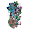
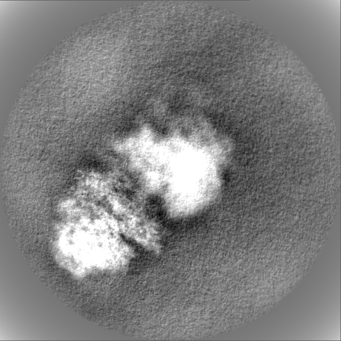
Journal: Proc Natl Acad Sci U S A / Year: 2021 Title: Structure of Gcn1 bound to stalled and colliding 80S ribosomes. Authors: Agnieszka A Pochopien / Bertrand Beckert / Sergo Kasvandik / Otto Berninghausen / Roland Beckmann / Tanel Tenson / Daniel N Wilson / Abstract: The Gcn pathway is conserved in all eukaryotes, including mammals such as humans, where it is a crucial part of the integrated stress response (ISR). Gcn1 serves as an essential effector protein for ...The Gcn pathway is conserved in all eukaryotes, including mammals such as humans, where it is a crucial part of the integrated stress response (ISR). Gcn1 serves as an essential effector protein for the kinase Gcn2, which in turn is activated by stalled ribosomes, leading to phosphorylation of eIF2 and a subsequent global repression of translation. The fine-tuning of this adaptive response is performed by the Rbg2/Gir2 complex, a negative regulator of Gcn2. Despite the wealth of available biochemical data, information on structures of Gcn proteins on the ribosome has remained elusive. Here we present a cryo-electron microscopy structure of the yeast Gcn1 protein in complex with stalled and colliding 80S ribosomes. Gcn1 interacts with both 80S ribosomes within the disome, such that the Gcn1 HEAT repeats span from the P-stalk region on the colliding ribosome to the P-stalk and the A-site region of the lead ribosome. The lead ribosome is stalled in a nonrotated state with peptidyl-tRNA in the A-site, uncharged tRNA in the P-site, eIF5A in the E-site, and Rbg2/Gir2 in the A-site factor binding region. By contrast, the colliding ribosome adopts a rotated state with peptidyl-tRNA in a hybrid A/P-site, uncharged-tRNA in the P/E-site, and Mbf1 bound adjacent to the mRNA entry channel on the 40S subunit. Collectively, our findings reveal the interaction mode of the Gcn2-activating protein Gcn1 with colliding ribosomes and provide insight into the regulation of Gcn2 activation. The binding of Gcn1 to a disome has important implications not only for the Gcn2-activated ISR, but also for the general ribosome-associated quality control pathways.
History
Deposition
Mar 3, 2021
-
Header (metadata) release
Apr 14, 2021
-
Map release
Apr 14, 2021
-
Update
Jul 9, 2025
-
Current status
Jul 9, 2025
Processing site: PDBe / Status: Released
-
Structure visualization
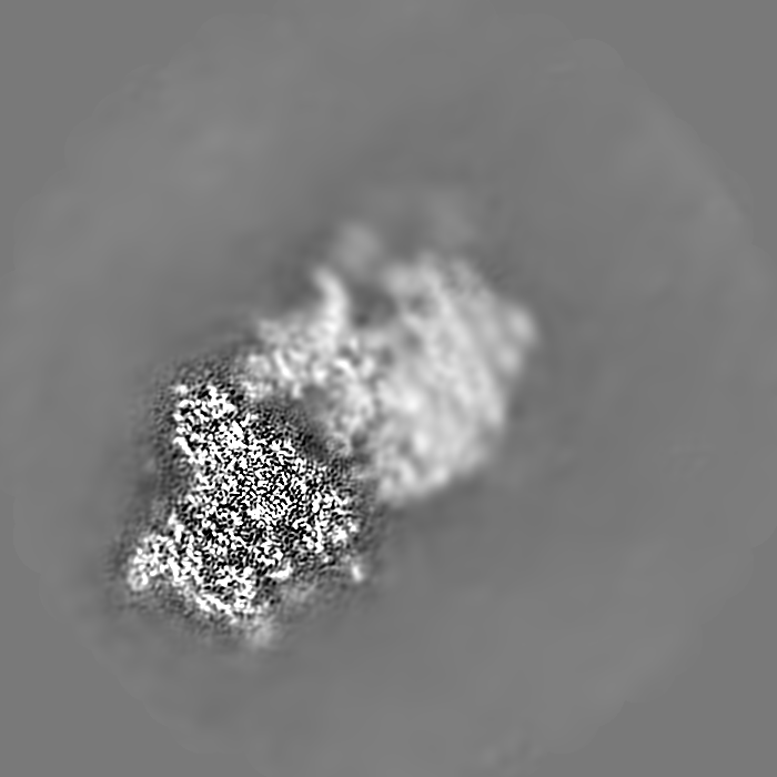
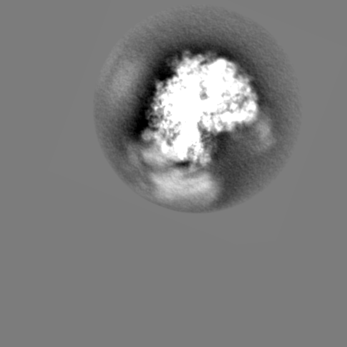
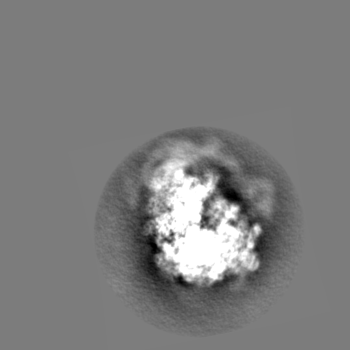
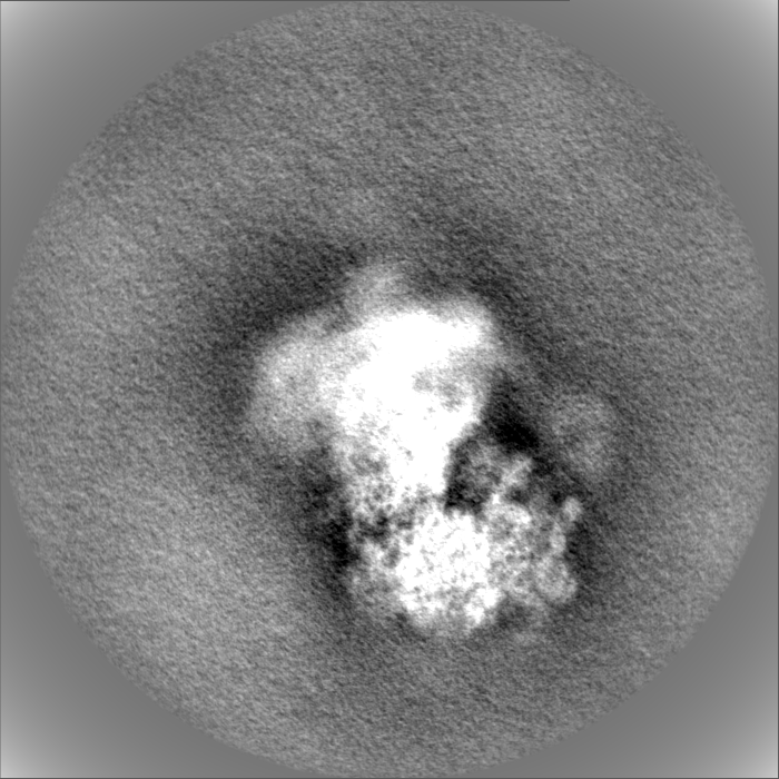
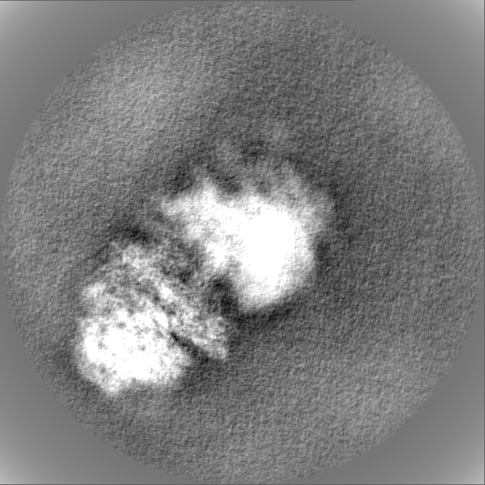
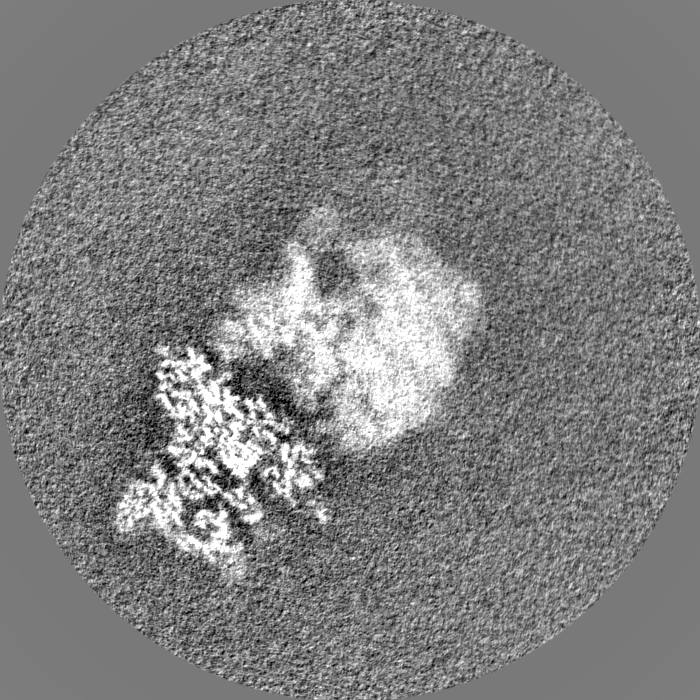
Movie
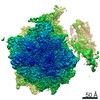
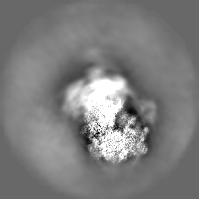
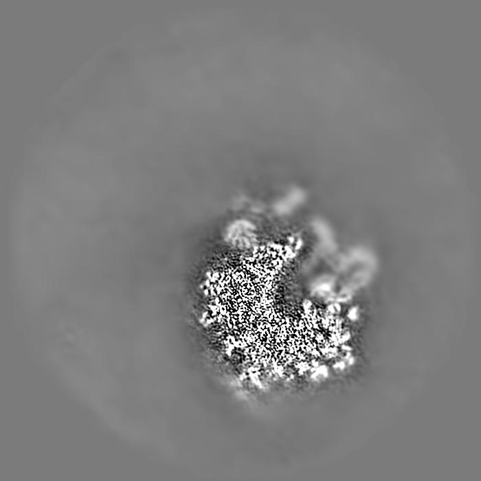
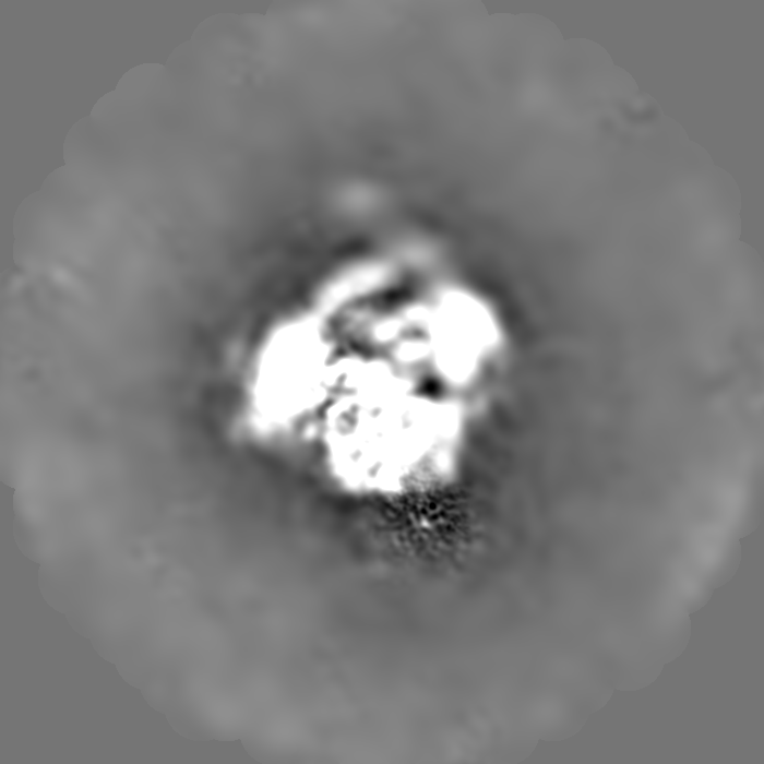
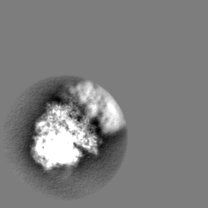
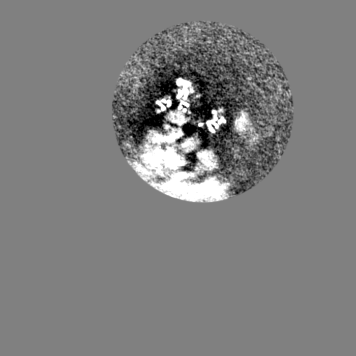
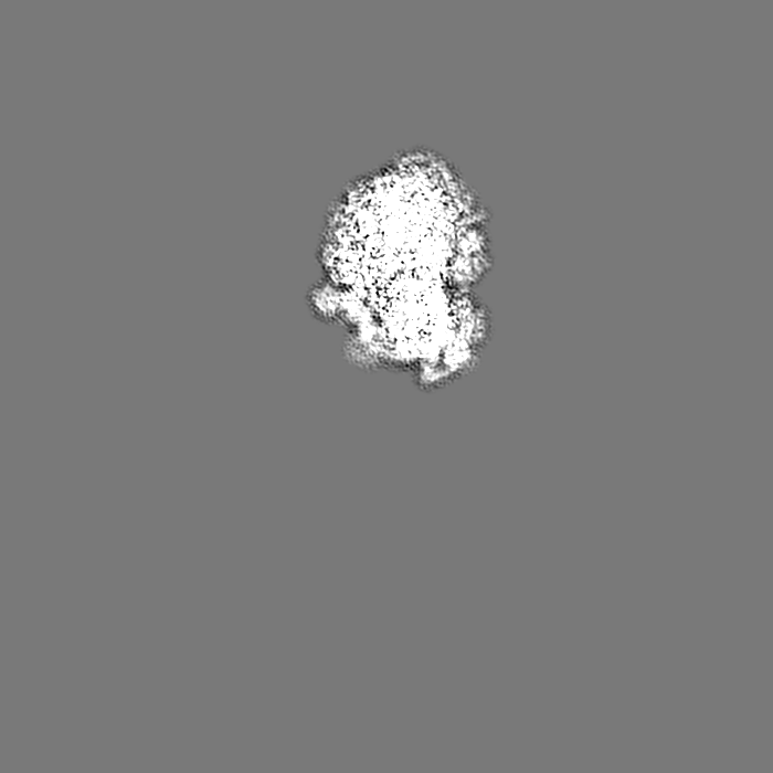
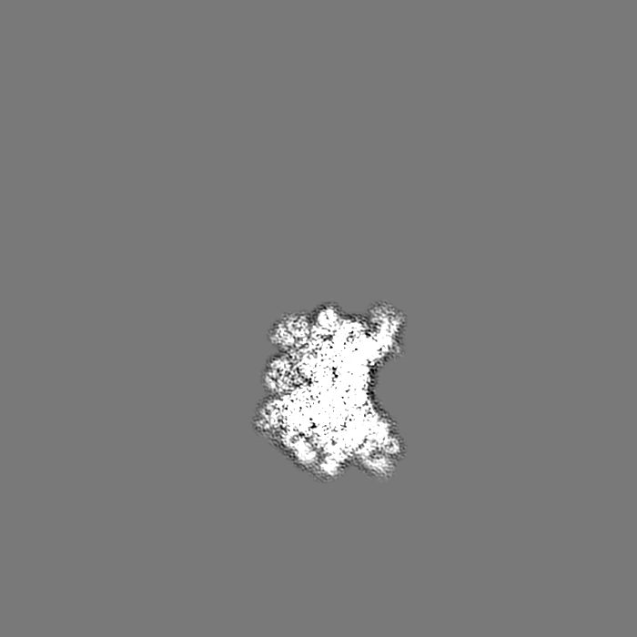
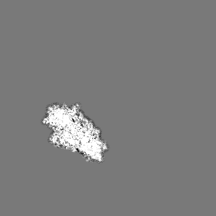
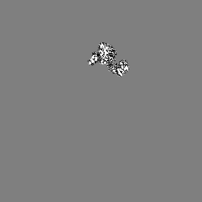
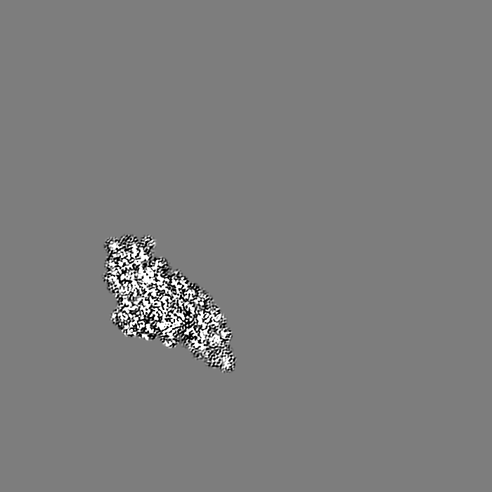
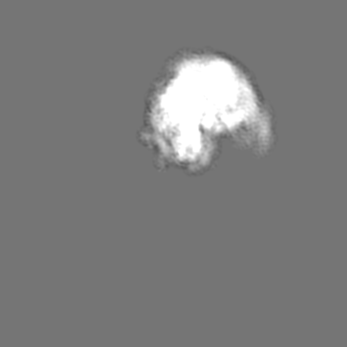
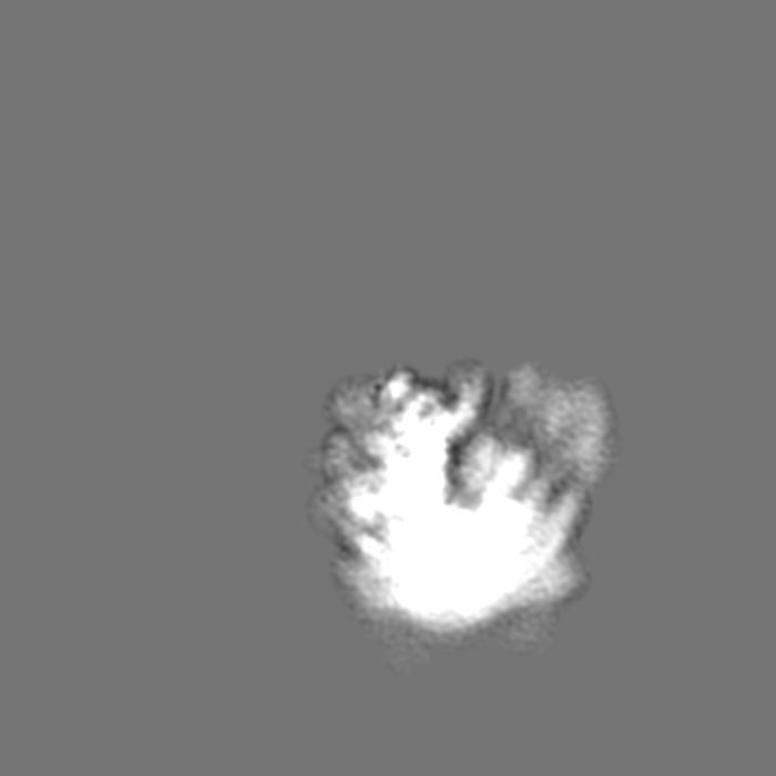
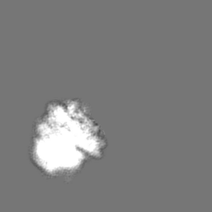
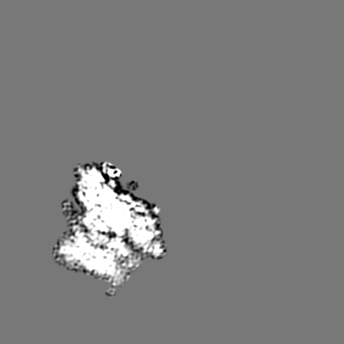
Surface view with section colored by density value
Name: 60S ribosomal protein L43-A / type: protein_or_peptide / ID: 82 / Number of copies: 1 / Enantiomer: LEVO
Source (natural)
Organism: Saccharomyces cerevisiae S288c (yeast)
Molecular weight
Theoretical: 9.981756 KDa
Sequence
String:
AKRTKKVGIT GKYGVRYGSS LRRQVKKLEI QQHARYDCSF CGKKTVKRGA AGIWTCSCCK KTVAGGAYTV STAAAATVRS TIRRLREMV EA
UniProtKB: Large ribosomal subunit protein eL43A
-
Experimental details
-
Structure determination
Method
cryo EM
Processing
single particle reconstruction
Aggregation state
particle
-
Sample preparation
Buffer
pH: 7.5
Grid
Model: Quantifoil R3/3 / Material: COPPER / Mesh: 300 / Support film - Material: CARBON / Support film - topology: CONTINUOUS / Support film - Film thickness: 300
Vitrification
Cryogen name: ETHANE-PROPANE
-
Electron microscopy
Microscope
FEI TITAN KRIOS
Image recording
Film or detector model: FEI FALCON II (4k x 4k) / Average electron dose: 2.5 e/Å2
Electron beam
Acceleration voltage: 300 kV / Electron source: FIELD EMISSION GUN
Electron optics
Illumination mode: SPOT SCAN / Imaging mode: BRIGHT FIELD
Experimental equipment
Model: Titan Krios / Image courtesy: FEI Company
+
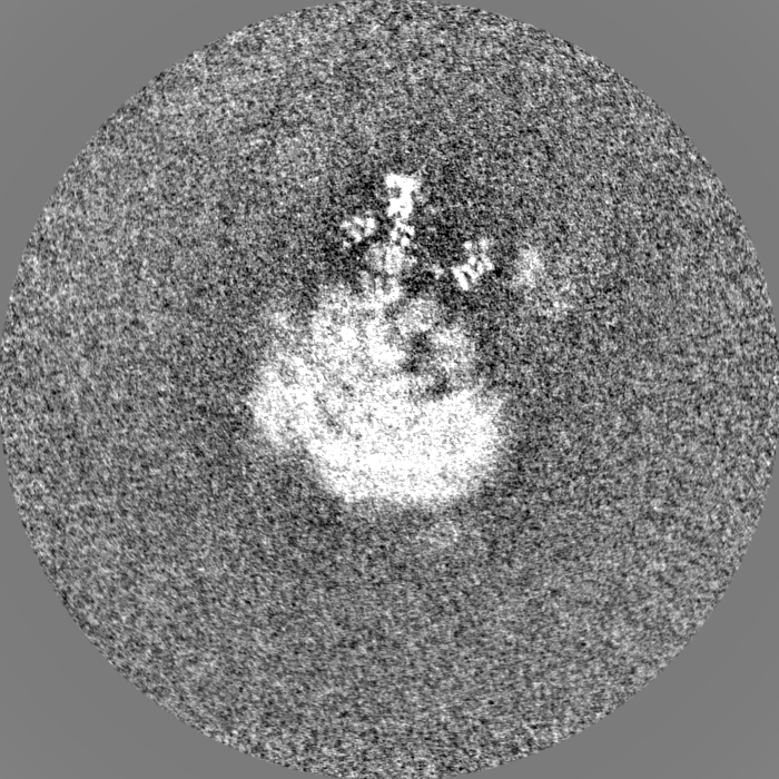
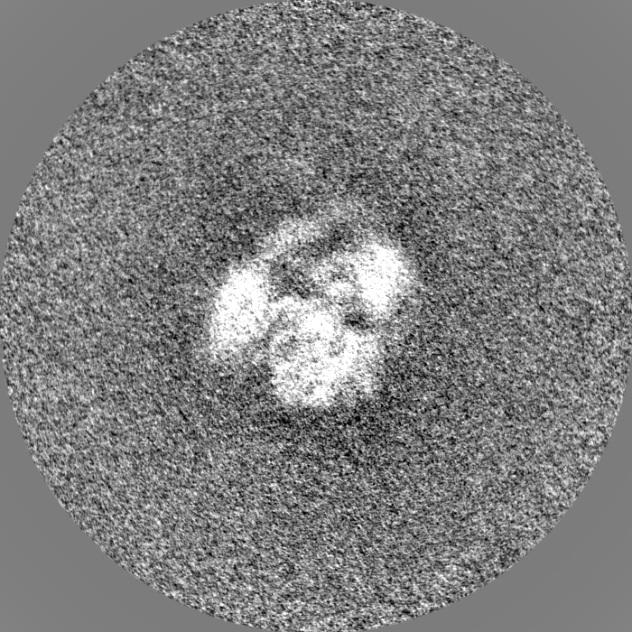
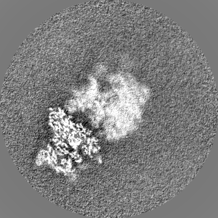
Image processing
CTF correction
Type: PHASE FLIPPING AND AMPLITUDE CORRECTION
Startup model
Type of model: PDB ENTRY
Final reconstruction
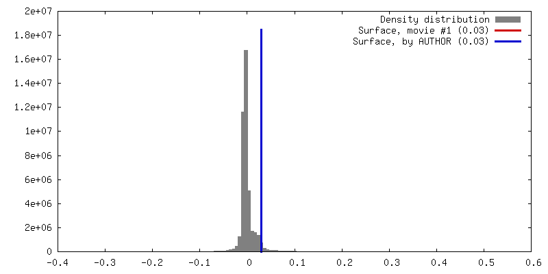
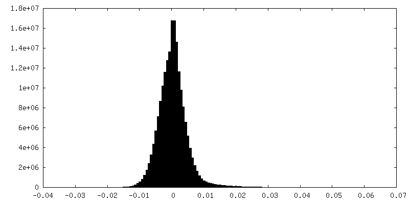
Applied symmetry - Point group: C1 (asymmetric) / Resolution.type: BY AUTHOR / Resolution: 4.36 Å / Resolution method: FSC 0.143 CUT-OFF / Software - Name: RELION (ver. 3.0) / Number images used: 30016
Initial angle assignment
Type: MAXIMUM LIKELIHOOD
Final angle assignment
Type: MAXIMUM LIKELIHOOD
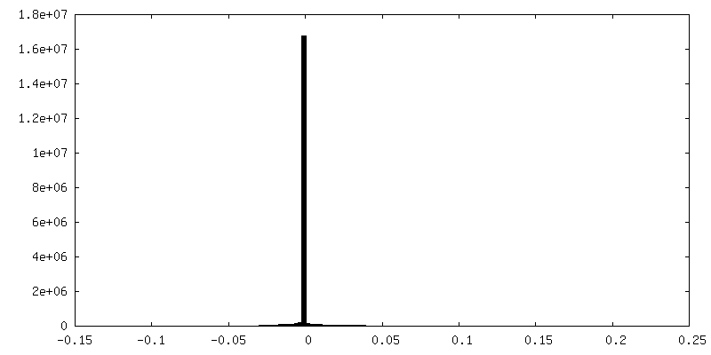
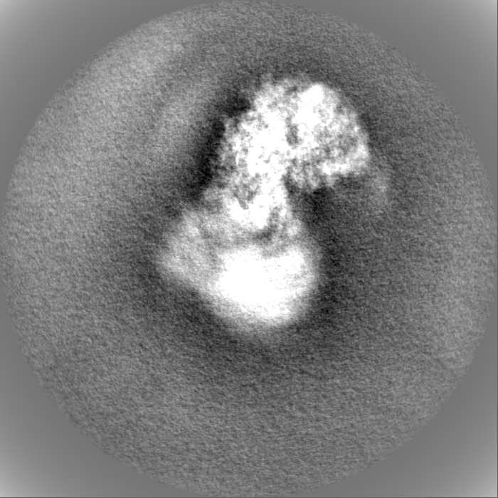
FSC plot (resolution estimation)
-
Atomic model buiding 1
Refinement
Space: REAL / Protocol: RIGID BODY FIT
Output model
PDB-7nrd: Structure of the yeast Gcn1 bound to a colliding stalled 80S ribosome with MBF1, A/P-tRNA and P/E-tRNA
+
About Yorodumi
-
News
-
Feb 9, 2022. New format data for meta-information of EMDB entries
New format data for meta-information of EMDB entries
Version 3 of the EMDB header file is now the official format.
The previous official version 1.9 will be removed from the archive.
In the structure databanks used in Yorodumi, some data are registered as the other names, "COVID-19 virus" and "2019-nCoV". Here are the details of the virus and the list of structure data.
Jan 31, 2019. EMDB accession codes are about to change! (news from PDBe EMDB page)
EMDB accession codes are about to change! (news from PDBe EMDB page)
The allocation of 4 digits for EMDB accession codes will soon come to an end. Whilst these codes will remain in use, new EMDB accession codes will include an additional digit and will expand incrementally as the available range of codes is exhausted. The current 4-digit format prefixed with “EMD-” (i.e. EMD-XXXX) will advance to a 5-digit format (i.e. EMD-XXXXX), and so on. It is currently estimated that the 4-digit codes will be depleted around Spring 2019, at which point the 5-digit format will come into force.
The EM Navigator/Yorodumi systems omit the EMD- prefix.
Related info.:Q: What is EMD? / ID/Accession-code notation in Yorodumi/EM Navigator
Yorodumi is a browser for structure data from EMDB, PDB, SASBDB, etc.
This page is also the successor to EM Navigator detail page, and also detail information page/front-end page for Omokage search.
The word "yorodu" (or yorozu) is an old Japanese word meaning "ten thousand". "mi" (miru) is to see.
Related info.:EMDB / PDB / SASBDB / Comparison of 3 databanks / Yorodumi Search / Aug 31, 2016. New EM Navigator & Yorodumi / Yorodumi Papers / Jmol/JSmol / Function and homology information / Changes in new EM Navigator and Yorodumi
 Movie
Movie Controller

 Yorodumi
 Open data
 Basic information
 Map data
 Sample
 Keywords
 Function and homology information
 Authors
 Germany, 2 items
  Citation
 Structure visualization
 Downloads & links
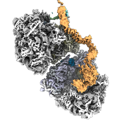 emd_12535.png
 http://ftp.pdbj.org/pub/emdb/structures/EMD-12535


 Z (Sec.)
 Y (Row.)
 X (Col.)


 Sample components
 Processing
 Electron microscopy
 FIELD EMISSION GUN

