- EMDB-30608: Cryo-EM Structure of the Prostaglandin E Receptor EP4 Coupled to ... -
+
Open data
ID or keywords:
Loading...
-
Basic information
Entry
Database: EMDB / ID: EMD-30608
Title
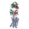
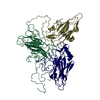
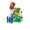
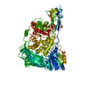
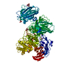
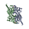
Cryo-EM Structure of the Prostaglandin E Receptor EP4 Coupled to G Protein
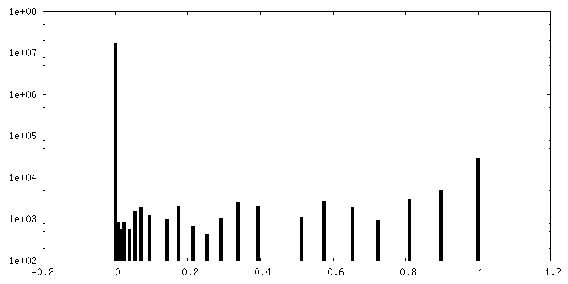
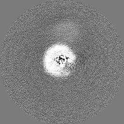
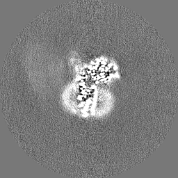
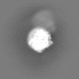
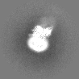
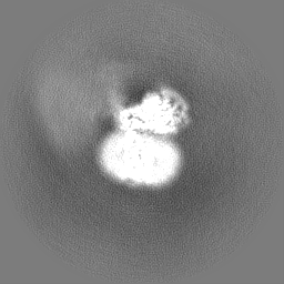
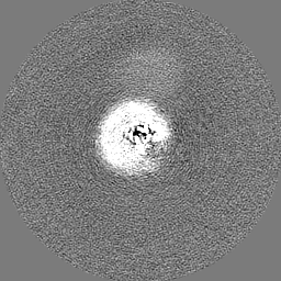
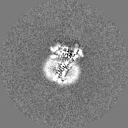
Map data
The unmasked final map
Sample
Complex: PGE2-EP4-Gs-Gbeta1-Ggamma2-Nb35 complex
Complex: Prostaglandin E2 receptor EP4 subtype,Guanine nucleotide-binding protein G(I)/G(S)/G(T) subunit beta-1,Guanine nucleotide-binding protein G(I)/G(S)/G(O) subunit gamma-2
Protein or peptide: Prostaglandin E2 receptor EP4 subtype,Prostaglandin E2 receptor EP4 subtype
Protein or peptide: Guanine nucleotide-binding protein G(I)/G(S)/G(T) subunit beta-1
Protein or peptide: Guanine nucleotide-binding protein G(I)/G(S)/G(O) subunit gamma-2
Complex: Guanine nucleotide-binding protein G(s) subunit alpha isoforms short
Protein or peptide: Guanine nucleotide-binding protein G(s) subunit alpha isoforms short,Guanine nucleotide-binding protein G(s) subunit alpha isoforms short,Guanine nucleotide-binding protein G(s) subunit alpha isoforms short
Japan Agency for Medical Research and Development (AMED)
JP20gm0910007
Japan
Japan Agency for Medical Research and Development (AMED)
JP20am0401020
Japan
Japan Agency for Medical Research and Development (AMED)
JP20ak0101103
Japan
Japan Agency for Medical Research and Development (AMED)
JP19am0101115
Japan
Citation
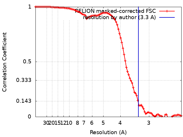
Journal: Structure / Year: 2021 Title: Cryo-EM Structure of the Prostaglandin E Receptor EP4 Coupled to G Protein. Authors: Shingo Nojima / Yoko Fujita / Kanako Terakado Kimura / Norimichi Nomura / Ryoji Suno / Kazushi Morimoto / Masaki Yamamoto / Takeshi Noda / So Iwata / Hideki Shigematsu / Takuya Kobayashi / Abstract: Prostaglandin E receptor EP4, a class A G protein-coupled receptor (GPCR), is a common drug target in various disorders, such as acute decompensated heart failure and ulcerative colitis. Here, we ...Prostaglandin E receptor EP4, a class A G protein-coupled receptor (GPCR), is a common drug target in various disorders, such as acute decompensated heart failure and ulcerative colitis. Here, we report the cryoelectron microscopy (cryo-EM) structure of the EP4-heterotrimeric G protein (Gs) complex with the endogenous ligand at a global resolution of 3.3 Å. In this structure, compared with that in the inactive EP4 structure, the sixth transmembrane domain is shifted outward on the intracellular side, although the shift is smaller than that in other class A GPCRs bound to Gs. Instead, the C-terminal helix of Gs is inserted toward TM2 of EP4, and the conserved C-terminal hook structure formsthe extended state. These structural features are formed by the conserved residues in prostanoid receptors (Phe54 and Trp327). These findings may be important for the thorough understanding of the G protein-binding mechanism of EP4 and other prostanoid receptors.
History
Deposition
Oct 5, 2020
-
Header (metadata) release
Nov 18, 2020
-
Map release
Nov 18, 2020
-
Update
Oct 23, 2024
-
Current status
Oct 23, 2024
Processing site: PDBj / Status: Released
-
Structure visualization
Movie
Surface view with section colored by density value
Complex: Prostaglandin E2 receptor EP4 subtype,Guanine nucleotide-binding protein G(I)/G(S)/G(T) subunit beta-1,Guanine nucleotide-binding protein G(I)/G(S)/G(O) subunit gamma-2
Protein or peptide: Prostaglandin E2 receptor EP4 subtype,Prostaglandin E2 receptor EP4 subtype
Protein or peptide: Guanine nucleotide-binding protein G(I)/G(S)/G(T) subunit beta-1
Protein or peptide: Guanine nucleotide-binding protein G(I)/G(S)/G(O) subunit gamma-2
Complex: Guanine nucleotide-binding protein G(s) subunit alpha isoforms short
Protein or peptide: Guanine nucleotide-binding protein G(s) subunit alpha isoforms short,Guanine nucleotide-binding protein G(s) subunit alpha isoforms short,Guanine nucleotide-binding protein G(s) subunit alpha isoforms short
UniProtKB: Guanine nucleotide-binding protein G(I)/G(S)/G(T) subunit beta-1
+
Macromolecule #3: Guanine nucleotide-binding protein G(I)/G(S)/G(O) subunit gamma-2
Macromolecule
Name: Guanine nucleotide-binding protein G(I)/G(S)/G(O) subunit gamma-2 type: protein_or_peptide / ID: 3 / Number of copies: 1 / Enantiomer: LEVO
Source (natural)
Organism: Homo sapiens (human)
Molecular weight
Theoretical: 7.861143 KDa
Recombinant expression
Organism: Spodoptera frugiperda (fall armyworm)
Sequence
String:
MASNNTASIA QARKLVEQLK MEANIDRIKV SKAAADLMAY CEAHAKEDPL LTPVPASENP FREKKFFCAI L
UniProtKB: Guanine nucleotide-binding protein G(I)/G(S)/G(O) subunit gamma-2
+
Macromolecule #4: Guanine nucleotide-binding protein G(s) subunit alpha isoforms sh...
Macromolecule
Name: Guanine nucleotide-binding protein G(s) subunit alpha isoforms short,Guanine nucleotide-binding protein G(s) subunit alpha isoforms short,Guanine nucleotide-binding protein G(s) subunit alpha isoforms short type: protein_or_peptide / ID: 4 Details: residues from 65 to 203, residues from 255 to 264 were deleted Number of copies: 1 / Enantiomer: LEVO
UniProtKB: Guanine nucleotide-binding protein G(s) subunit alpha isoforms short, Guanine nucleotide-binding protein G(s) subunit alpha isoforms short, Guanine nucleotide-binding protein G(s) subunit alpha isoforms short
+
Macromolecule #5: nanobody Nb35
Macromolecule
Name: nanobody Nb35 / type: protein_or_peptide / ID: 5 Details: The C-terminal "ENLYFQ" of sample sequence is a cleavaged TEV protease recognition sequence. Number of copies: 1 / Enantiomer: LEVO
Model: Quantifoil R0.6/1 / Material: COPPER / Mesh: 300 / Support film - Material: CARBON / Support film - topology: HOLEY ARRAY / Pretreatment - Type: GLOW DISCHARGE / Pretreatment - Time: 10 sec. / Pretreatment - Atmosphere: AIR / Pretreatment - Pressure: 0.007 kPa / Details: 10 mA
Vitrification
Cryogen name: ETHANE / Chamber humidity: 100 % / Chamber temperature: 281 K / Instrument: FEI VITROBOT MARK IV / Details: Blot Force 10, Blot Time 3.5 sec, 3 microL apply.
-
Electron microscopy
Microscope
FEI TITAN KRIOS
Specialist optics
Energy filter - Name: GIF Bioquantum / Energy filter - Slit width: 25 eV
Image recording
Film or detector model: GATAN K3 (6k x 4k) / Digitization - Dimensions - Width: 4092 pixel / Digitization - Dimensions - Height: 5760 pixel / Number grids imaged: 1 / Number real images: 5743 / Average exposure time: 1.83 sec. / Average electron dose: 50.0 e/Å2
Electron beam
Acceleration voltage: 300 kV / Electron source: FIELD EMISSION GUN
In the structure databanks used in Yorodumi, some data are registered as the other names, "COVID-19 virus" and "2019-nCoV". Here are the details of the virus and the list of structure data.
Jan 31, 2019. EMDB accession codes are about to change! (news from PDBe EMDB page)
EMDB accession codes are about to change! (news from PDBe EMDB page)
The allocation of 4 digits for EMDB accession codes will soon come to an end. Whilst these codes will remain in use, new EMDB accession codes will include an additional digit and will expand incrementally as the available range of codes is exhausted. The current 4-digit format prefixed with “EMD-” (i.e. EMD-XXXX) will advance to a 5-digit format (i.e. EMD-XXXXX), and so on. It is currently estimated that the 4-digit codes will be depleted around Spring 2019, at which point the 5-digit format will come into force.
The EM Navigator/Yorodumi systems omit the EMD- prefix.
Related info.:Q: What is EMD? / ID/Accession-code notation in Yorodumi/EM Navigator
Yorodumi is a browser for structure data from EMDB, PDB, SASBDB, etc.
This page is also the successor to EM Navigator detail page, and also detail information page/front-end page for Omokage search.
The word "yorodu" (or yorozu) is an old Japanese word meaning "ten thousand". "mi" (miru) is to see.
Related info.:EMDB / PDB / SASBDB / Comparison of 3 databanks / Yorodumi Search / Aug 31, 2016. New EM Navigator & Yorodumi / Yorodumi Papers / Jmol/JSmol / Function and homology information / Changes in new EM Navigator and Yorodumi
 Movie
Movie Controller

 Yorodumi
 Open data
 Basic information
 Map data
 Sample
 Keywords
 Function and homology information
 Homo sapiens (human) /
 Authors
 Japan, 5 items
  Citation
 Structure visualization
 Downloads & links
 emd_30608.png
 http://ftp.pdbj.org/pub/emdb/structures/EMD-30608
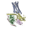


 Z (Sec.)
 Y (Row.)
 X (Col.)


 Sample components
 Spodoptera frugiperda (fall armyworm)
 Processing
 Electron microscopy
 FIELD EMISSION GUN