 Movie
Movie Controller

[English] 日本語
 Yorodumi
- PDB-6gdg: Cryo-EM structure of the adenosine A2A receptor bound to a miniGs... -
+ Open data

- Basic information
| Entry | Database: PDB / ID: 6gdg | |||||||||
|---|---|---|---|---|---|---|---|---|---|---|
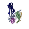
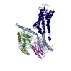
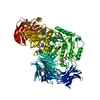
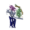
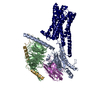
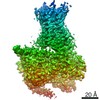
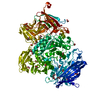
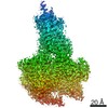
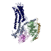
| Title | Cryo-EM structure of the adenosine A2A receptor bound to a miniGs heterotrimer | |||||||||
 Components Components |
| |||||||||
 Keywords Keywords | MEMBRANE PROTEIN / G-protein coupled receptor / adenosine / GPCR / A2A receptor | |||||||||
| Function / homology |  Function and homology information Function and homology informationregulation of norepinephrine secretion / negative regulation of alpha-beta T cell activation / positive regulation of circadian sleep/wake cycle, sleep / Adenosine P1 receptors / positive regulation of acetylcholine secretion, neurotransmission / G protein-coupled adenosine receptor activity / response to purine-containing compound / G protein-coupled adenosine receptor signaling pathway / NGF-independant TRKA activation / Surfactant metabolism ...regulation of norepinephrine secretion / negative regulation of alpha-beta T cell activation / positive regulation of circadian sleep/wake cycle, sleep / Adenosine P1 receptors / positive regulation of acetylcholine secretion, neurotransmission / G protein-coupled adenosine receptor activity / response to purine-containing compound / G protein-coupled adenosine receptor signaling pathway / NGF-independant TRKA activation / Surfactant metabolism / synaptic transmission, dopaminergic / type 5 metabotropic glutamate receptor binding / negative regulation of vascular permeability / synaptic transmission, cholinergic / intermediate filament / presynaptic active zone / positive regulation of urine volume / response to caffeine / blood circulation / sensory perception / positive regulation of glutamate secretion / DNA polymerase processivity factor activity / eating behavior / inhibitory postsynaptic potential / regulation of calcium ion transport / adenylate cyclase-activating G protein-coupled bile acid receptor signaling pathway / adenylate cyclase-activating serotonin receptor signaling pathway / protein-disulfide reductase activity / regulation of skeletal muscle contraction / alpha-actinin binding / PKA activation in glucagon signalling / hair follicle placode formation / developmental growth / intracellular transport / asymmetric synapse / axolemma / membrane depolarization / D1 dopamine receptor binding / cellular defense response / prepulse inhibition / vascular endothelial cell response to laminar fluid shear stress / phagocytosis / renal water homeostasis / Hedgehog 'off' state / activation of adenylate cyclase activity / adenylate cyclase-activating adrenergic receptor signaling pathway / cellular response to acidic pH / neuron projection morphogenesis / positive regulation of synaptic transmission, glutamatergic / cellular response to glucagon stimulus / cell redox homeostasis / astrocyte activation / intracellular glucose homeostasis / presynaptic modulation of chemical synaptic transmission / positive regulation of insulin secretion involved in cellular response to glucose stimulus / adenylate cyclase activator activity / trans-Golgi network membrane / positive regulation of long-term synaptic potentiation / positive regulation of synaptic transmission, GABAergic / central nervous system development / positive regulation of protein secretion / response to amphetamine / regulation of mitochondrial membrane potential / positive regulation of apoptotic signaling pathway / negative regulation of inflammatory response to antigenic stimulus / apoptotic signaling pathway / synaptic transmission, glutamatergic / response to prostaglandin E / excitatory postsynaptic potential / locomotory behavior / bone development / platelet aggregation / negative regulation of inflammatory response / cognition / vasodilation / G-protein beta/gamma-subunit complex binding / adenylate cyclase-modulating G protein-coupled receptor signaling pathway / blood coagulation / positive regulation of insulin secretion / Olfactory Signaling Pathway / Activation of the phototransduction cascade / G protein-coupled acetylcholine receptor signaling pathway / G beta:gamma signalling through PLC beta / Presynaptic function of Kainate receptors / Thromboxane signalling through TP receptor / sensory perception of smell / Activation of G protein gated Potassium channels / Inhibition of voltage gated Ca2+ channels via Gbeta/gamma subunits / G-protein activation / Glucagon signaling in metabolic regulation / Prostacyclin signalling through prostacyclin receptor / G beta:gamma signalling through CDC42 / Synthesis, secretion, and inactivation of Glucagon-like Peptide-1 (GLP-1) / G beta:gamma signalling through BTK / photoreceptor disc membrane / ADP signalling through P2Y purinoceptor 12 / Glucagon-type ligand receptors / Sensory perception of sweet, bitter, and umami (glutamate) taste / Adrenaline,noradrenaline inhibits insulin secretion / Vasopressin regulates renal water homeostasis via Aquaporins Similarity search - Function | |||||||||
| Biological species |   Homo sapiens (human) Homo sapiens (human) | |||||||||
| Method | ELECTRON MICROSCOPY / single particle reconstruction / cryo EM / Resolution: 4.11 Å | |||||||||
 Authors Authors | Garcia-Nafria, J. / Lee, Y. | |||||||||
| Funding support |  United Kingdom, 2items United Kingdom, 2items
| |||||||||
 Citation Citation | Journal: Elife / Year: 2018 Title: Cryo-EM structure of the adenosine A receptor coupled to an engineered heterotrimeric G protein. Authors: Javier García-Nafría / Yang Lee / Xiaochen Bai / Byron Carpenter / Christopher G Tate / Abstract: The adenosine A receptor (AR) is a prototypical G protein-coupled receptor (GPCR) that couples to the heterotrimeric G protein G. Here, we determine the structure by electron cryo-microscopy (cryo-EM) ...The adenosine A receptor (AR) is a prototypical G protein-coupled receptor (GPCR) that couples to the heterotrimeric G protein G. Here, we determine the structure by electron cryo-microscopy (cryo-EM) of AR at pH 7.5 bound to the small molecule agonist NECA and coupled to an engineered heterotrimeric G protein, which contains mini-G, the βγ subunits and nanobody Nb35. Most regions of the complex have a resolution of ~3.8 Å or better. Comparison with the 3.4 Å resolution crystal structure shows that the receptor and mini-G are virtually identical and that the density of the side chains and ligand are of comparable quality. However, the cryo-EM density map also indicates regions that are flexible in comparison to the crystal structures, which unexpectedly includes regions in the ligand binding pocket. In addition, an interaction between intracellular loop 1 of the receptor and the β subunit of the G protein was observed. | |||||||||
| History |
|
- Structure visualization
| Movie |
Movie viewer |
|---|---|
| Structure viewer | Molecule: MolmilJmol/JSmol |
- Downloads & links
-Download
| PDBx/mmCIF format | 6gdg.cif.gz | 211.1 KB | Display | PDBx/mmCIF format |
|---|---|---|---|---|
| PDB format | pdb6gdg.ent.gz | 155 KB | Display | PDB format |
| PDBx/mmJSON format | 6gdg.json.gz | Tree view | PDBx/mmJSON format | |
| Others |  Other downloads Other downloads |
-Validation report
| Arichive directory | https://data.pdbj.org/pub/pdb/validation_reports/gd/6gdgftp://data.pdbj.org/pub/pdb/validation_reports/gd/6gdg | HTTPS FTP |
|---|
-Related structure data
| Related structure data |  4390MC M: map data used to model this data C: citing same article ( |
|---|---|
| Similar structure data | |
| EM raw data | EMPIAR-10309 (Title: Cryo-EM structure of the adenosine A2A receptor coupled to an engineered heterotrimeric G protein Data size: 1.1 TB Data #1: Unaligned multi-frame micrographs [micrographs - multiframe] Data #2: Micelle subtracted particles [picked particles - multiframe - processed] Data #3: Particles before subtraction [picked particles - multiframe - processed]) |

-Links
 PDBj
PDBj


- Assembly
| Deposited unit | 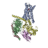
|
|---|---|
| 1 |
|
-Components
-Guanine nucleotide-binding protein ... , 3 types, 3 molecules BCD
| #2: Protein | Mass: 37285.734 Da / Num. of mol.: 1 Source method: isolated from a genetically manipulated source Source: (gene. exp.) Homo sapiens (human) / Gene: GNB1 / Production host:  Trichoplusia ni (cabbage looper) / References: UniProt: P62873 Trichoplusia ni (cabbage looper) / References: UniProt: P62873 |
|---|---|
| #3: Protein | Mass: 7845.078 Da / Num. of mol.: 1 Source method: isolated from a genetically manipulated source Source: (gene. exp.) Homo sapiens (human) / Gene: GNG2 / Production host: Trichoplusia ni (cabbage looper) / References: UniProt: P59768 |
| #4: Protein | Mass: 28907.684 Da / Num. of mol.: 1 Source method: isolated from a genetically manipulated source Source: (gene. exp.) Homo sapiens (human) / Gene: GNAS, GNAS1, GSP / Production host: Trichoplusia ni (cabbage looper) / References: UniProt: P63092 |
-Protein / Antibody / Non-polymers , 3 types, 3 molecules AE
| #1: Protein | Mass: 52909.785 Da / Num. of mol.: 1 / Fragment: UNP residues 37-144,UNP residues 8-316 Source method: isolated from a genetically manipulated source Source: (gene. exp.) Homo sapiens (human)Gene: trxA, ADORA2A, ADORA2 / Production host: Trichoplusia ni (cabbage looper)References: UniProt: Q14F07, UniProt: P29274, UniProt: P0AA25*PLUS |
|---|---|
| #5: Antibody | Mass: 16926.076 Da / Num. of mol.: 1 Source method: isolated from a genetically manipulated source Source: (gene. exp.) |
| #6: Chemical | ChemComp-NEC /  Mass: 308.293 Da / Num. of mol.: 1 / Source method: obtained synthetically / Formula: C12H16N6O4 Mass: 308.293 Da / Num. of mol.: 1 / Source method: obtained synthetically / Formula: C12H16N6O4 |
-Details
| Has protein modification | Y |
|---|
-Experimental details
-Experiment
| Experiment | Method: ELECTRON MICROSCOPY |
|---|---|
| EM experiment | Aggregation state: PARTICLE / 3D reconstruction method: single particle reconstruction |
- Sample preparation
| Component |
| ||||||||||||||||||||||||||||||
|---|---|---|---|---|---|---|---|---|---|---|---|---|---|---|---|---|---|---|---|---|---|---|---|---|---|---|---|---|---|---|---|
| Molecular weight | Value: 0.137 MDa / Experimental value: NO | ||||||||||||||||||||||||||||||
| Source (natural) |
| ||||||||||||||||||||||||||||||
| Source (recombinant) |
| ||||||||||||||||||||||||||||||
| Buffer solution | pH: 7.5 | ||||||||||||||||||||||||||||||
| Specimen | Conc.: 1 mg/ml / Embedding applied: NO / Shadowing applied: NO / Staining applied: NO / Vitrification applied: YES | ||||||||||||||||||||||||||||||
| Specimen support | Grid material: GOLD / Grid mesh size: 300 divisions/in. / Grid type: Quantifoil R1.2/1.3 | ||||||||||||||||||||||||||||||
| Vitrification | Instrument: FEI VITROBOT MARK IV / Cryogen name: ETHANE / Humidity: 100 % / Chamber temperature: 277.15 K |
- Electron microscopy imaging
| Experimental equipment |  Model: Titan Krios / Image courtesy: FEI Company |
|---|---|
| Microscopy | Model: FEI TITAN KRIOS |
| Electron gun | Electron source:  FIELD EMISSION GUN / Accelerating voltage: 300 kV / Illumination mode: FLOOD BEAM FIELD EMISSION GUN / Accelerating voltage: 300 kV / Illumination mode: FLOOD BEAM |
| Electron lens | Mode: BRIGHT FIELD / Cs: 2.7 mm / C2 aperture diameter: 50 µm / Alignment procedure: COMA FREE |
| Specimen holder | Cryogen: NITROGEN / Specimen holder model: FEI TITAN KRIOS AUTOGRID HOLDER |
| Image recording | Average exposure time: 60 sec. / Electron dose: 30 e/Å2 / Detector mode: COUNTING / Film or detector model: FEI FALCON III (4k x 4k) |
- Processing
| EM software |
| ||||||||||||||||||||||||||||||||||||||||
|---|---|---|---|---|---|---|---|---|---|---|---|---|---|---|---|---|---|---|---|---|---|---|---|---|---|---|---|---|---|---|---|---|---|---|---|---|---|---|---|---|---|
| CTF correction | Type: PHASE FLIPPING AND AMPLITUDE CORRECTION | ||||||||||||||||||||||||||||||||||||||||
| Symmetry | Point symmetry: C1 (asymmetric) | ||||||||||||||||||||||||||||||||||||||||
| 3D reconstruction | Resolution: 4.11 Å / Resolution method: FSC 0.143 CUT-OFF / Num. of particles: 128002 / Algorithm: FOURIER SPACE / Symmetry type: POINT | ||||||||||||||||||||||||||||||||||||||||
| Atomic model building | Protocol: FLEXIBLE FIT / Space: RECIPROCAL |
